Skin is composed of the epidermis, dermis, and adnexal structures. The epidermis is composed of 4 layers—the stratums basale, spinosum, granulosum, and corneum. The dermis is divided into a superficial papillary dermis and deeper reticular dermis. Collagen and elastin within the reticular dermis are responsible for skin tensile strength and elasticity, respectively. The 2 most common kinds of nonmelanoma skin cancers are basal cell and squamous cell carcinoma. Both are caused by a host of environmental and genetic factors, although UV light exposure is the single greatest predisposing factor.
Key points
- •
Skin is composed of 2 layers: the epidermis and dermis. Beneath it lays the hypodermis or subcutaneous tissue.
- •
The epidermis is composed of 4 layers: the stratum basale, spinosum, granulosum, and corneum.
- •
The dermis is composed of a thin, looser papillary dermis and a thicker, denser reticular dermis.
- •
The most commonly diagnosed nonmelanoma skin cancers are basal cell carcinoma and squamous cell carcinoma.
- •
Basal cell and squamous cell carcinomas are caused by a host of environmental and genetic factors.
Skin consists of 2 basic layers, the epidermis and dermis. The epidermis is primarily composed of keratinocytes but also contains melanocytes, Langerhans cells, and Merkel cells. It is divided into 4 layers or strata that are traversed by skin appendages such as pilosebaceous units and sweat glands. The dermis is divided into a papillary and reticular layer. Within the dermis resides the skin’s neurovascular supply. The subcutaneous tissue beneath the skin contains the superficial fascia and subcutaneous fat.
Epidermis
The epidermis is the outermost layer of skin ( Fig. 1 ). It is responsible for skin color, texture, and moisture. Epidermal thickness is relatively constant throughout the head and neck region. The primary cell type within the epidermis is the keratinocyte, and the 4 epidermal layers represent the maturation of keratinocytes from the deep to superficial layer. This process of keratinization allows for the development of keratin, a protein filament.
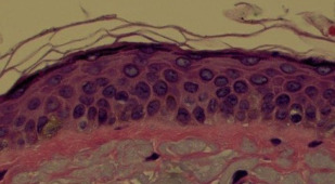
The deepest layer of the epidermis is the stratum basale, or basal layer. It is composed of stem cells called basal cells. The basal layer is often 1 cell thick, but can be 2 or 3 cells thick. Basal cells divide to form keratinocytes, which then begin migrating superficially.
The next layer is the spinous layer or stratum spinosum. Keratinocytes in this layer form intercellular attachments via protein channels called desmosomes. The attachments are responsible for this layer’s spiny appearance beneath the microscope. Lipid-containing lamellar granules first become visible here within the keratinocytes.
Keratinocytes then migrate to the granular layer or stratum granulosum, so named for the visible keratohyalin granules. Fillagrin forms within these granules from its precursor protein, profillagrin. Keratin filaments then begin to aggregate into complex structures via fillagrin.
Cells within the granular layer gradually lose their organelles and become more compact. They form the outermost epidermal layer, the stratum corneum. Here, keratinization is completed. The keratinocytes attach to one another via desmosomes in a bricklike pattern and are surrounded by lipids secreted from the lamellar granules. This construct is responsible for the skin’s function as a both a protective and a moisture-control barrier.
Cells
The keratinocyte is the primary cell type within the epidermis, but other cell types reside there as well. Melanocytes are confined to the basal layer. Their primary function is to produce melanin, a pigment that protects cellular nuclei from UV radiation-induced injury. Melanin-containing vesicles called melanosomes are secreted from the melanocyte dendritic processes and taken up by adjacent keratinocytes. The melanin pigment is then distributed over the nuclei to maximize the protection of DNA. Variations in skin color are not due to the number of melanocytes but rather to their activity level and volume of melanin production.
Langerhans cells are antigen processing and presenting cells found in the stratum spinosum, stratum granularum, and the dermis. Electron microscopy reveals racket-shaped granules called Birbeck granules. Langerhans cells have dendritic processes similar to melanocytes. Langerhans cell numbers decrease with UV radiation exposure, and the consequent decrease in skin immunologic activity may create a more permissive environment for carcinoma development.
Another epidermal cell type is the Merkel cell. Merkel cells reside in the basal layer and contain secretory granules whose contents are similar to those in other neuroendocrine cells. Groups of Merkel cells associated with peripheral nerve endings form specialized structures called tactile discs, which most likely facilitate fine sensation. They are predictably dense within and around highly sensitive locations and structures such as the lips, oral cavity, and hair follicles.
Dermal-Epidermal Junction
The basal layer of the epidermis is connected to the dermis below by a basement membrane called the dermal-epidermal junction. Two distinct layers of this junction are visible on electron microscopy. The more superficial layer, the lamina lucida, is composed of anchor filaments connecting hemidesmosomes within the basal cell plasma membrane to the deeper, more compact layer known as the lamina densa. The lamina densa is connected to the underlying dermis via collagen-anchoring fibrils.
Epidermis
The epidermis is the outermost layer of skin ( Fig. 1 ). It is responsible for skin color, texture, and moisture. Epidermal thickness is relatively constant throughout the head and neck region. The primary cell type within the epidermis is the keratinocyte, and the 4 epidermal layers represent the maturation of keratinocytes from the deep to superficial layer. This process of keratinization allows for the development of keratin, a protein filament.
The deepest layer of the epidermis is the stratum basale, or basal layer. It is composed of stem cells called basal cells. The basal layer is often 1 cell thick, but can be 2 or 3 cells thick. Basal cells divide to form keratinocytes, which then begin migrating superficially.
The next layer is the spinous layer or stratum spinosum. Keratinocytes in this layer form intercellular attachments via protein channels called desmosomes. The attachments are responsible for this layer’s spiny appearance beneath the microscope. Lipid-containing lamellar granules first become visible here within the keratinocytes.
Keratinocytes then migrate to the granular layer or stratum granulosum, so named for the visible keratohyalin granules. Fillagrin forms within these granules from its precursor protein, profillagrin. Keratin filaments then begin to aggregate into complex structures via fillagrin.
Cells within the granular layer gradually lose their organelles and become more compact. They form the outermost epidermal layer, the stratum corneum. Here, keratinization is completed. The keratinocytes attach to one another via desmosomes in a bricklike pattern and are surrounded by lipids secreted from the lamellar granules. This construct is responsible for the skin’s function as a both a protective and a moisture-control barrier.
Cells
The keratinocyte is the primary cell type within the epidermis, but other cell types reside there as well. Melanocytes are confined to the basal layer. Their primary function is to produce melanin, a pigment that protects cellular nuclei from UV radiation-induced injury. Melanin-containing vesicles called melanosomes are secreted from the melanocyte dendritic processes and taken up by adjacent keratinocytes. The melanin pigment is then distributed over the nuclei to maximize the protection of DNA. Variations in skin color are not due to the number of melanocytes but rather to their activity level and volume of melanin production.
Langerhans cells are antigen processing and presenting cells found in the stratum spinosum, stratum granularum, and the dermis. Electron microscopy reveals racket-shaped granules called Birbeck granules. Langerhans cells have dendritic processes similar to melanocytes. Langerhans cell numbers decrease with UV radiation exposure, and the consequent decrease in skin immunologic activity may create a more permissive environment for carcinoma development.
Another epidermal cell type is the Merkel cell. Merkel cells reside in the basal layer and contain secretory granules whose contents are similar to those in other neuroendocrine cells. Groups of Merkel cells associated with peripheral nerve endings form specialized structures called tactile discs, which most likely facilitate fine sensation. They are predictably dense within and around highly sensitive locations and structures such as the lips, oral cavity, and hair follicles.
Dermal-Epidermal Junction
The basal layer of the epidermis is connected to the dermis below by a basement membrane called the dermal-epidermal junction. Two distinct layers of this junction are visible on electron microscopy. The more superficial layer, the lamina lucida, is composed of anchor filaments connecting hemidesmosomes within the basal cell plasma membrane to the deeper, more compact layer known as the lamina densa. The lamina densa is connected to the underlying dermis via collagen-anchoring fibrils.
Related posts:


Stay updated, free articles. Join our Telegram channel

Full access? Get Clinical Tree


