Important advances in fibroblastic and fibrohistiocytic tumors relevant to dermatologists and dermatopathologists include (1) recognition that myxofibrosarcoma is a distinct entity that frequently arises in skin; (2) CD10 is sensitive but not specific atypical fibroxanthoma; (3) neurothekeomas lacking S100 expression are probably fibrohistiocytic/fibroblastic tumors, whereas S100+ myxoid variants are better classified as nerve sheath myxomas; (4) the recognition of a primary cutaneous variant of solitary fibrous tumor; (5) thelimitations of b-catenin immunohistochemistry in desmoid tumors; and (6) the prognostic utility of clinical and histopathologic variables in dermatofibrosarcoma protuberans, and the effects of imatinib mesylate therapy.
- •
The recognition that myxofibrosarcoma is a distinct entity that often presents in skin.
- •
CD10 is not specific for atypical fibroxanthoma (AFX).
- •
Evidence that ‘neurothekeomas’ are likely fibroblastic/fibrohistiocytic tumors, while S100+ myxoid variants are better classified as ‘nerve sheath myxomas’.
- •
The recognition of primary cutaneous solitary fibrous tumors.
- •
The limitations of beta-catenin immunohistochemistry in desmoid tumors.
- •
Prognostic utility of clinical and histopathologic features in dermatofibrosarcoma protuberans, and the effects of imatinibmesylate therapy.
Introduction
Recent advances in fibroblastic and fibrohistiocytic tumors have led to the following conclusions: (1) myxofibrosarcoma is a distinct entity with a propensity to present in the skin; (2) CD10 is not at all specifc for atypical fibroxanthoma; (3) nerve sheath myxomas are distinct from neurothekeomas; (4) solitary fibrous tumors may be primary to the skin, and most appear to be indolent; (5) beta-catenin immunohistochemistry is of only limited value in the diagnosis of desmoid tumors; and (6) metastasis remains the only relevant predictor of survival in dermatofibrosarcoma protuberans; imatinib mesylate therapy may complicate margin interpretation.
Myxofibrosarcoma: a distinctive sarcoma that commonly arises in skin
The myxofibrosarcoma is not new. What is new is its recognition as a distinctive sarcoma that commonly presents in the skin. Angervall and colleagues characterized myxofibrosarcomas more than 35 years ago, but the study was largely ignored and most pathologists considered this tumor merely a myxoid variant of so-called malignant fibrous histiocytoma (MFH).
During the past decade, however, several sarcoma experts have challenged the concept of fibrohistiocytic tumors in general, because no common line of fibrohistiocytic differentiation has been defined. Instead, several studies showed that many tumors traditionally classified as MFH have genetic aberrations commonly encountered in other high-grade sarcomas, such as liposarcoma or leiomyosarcoma. Some experts believe that with diligent study, most high-grade sarcomas can be classified as something other than MFH. As a result, some recommend abandoning the term MFH in favor of “undifferentiated pleomorphic sarcoma” (UPS) for these tumors.
Much of the debate is academic, but a practical by-product is the increasing awareness that myxofibrosarcoma is not myxoid MFH. Myxofibrosarcomas and MFH/UPS actually differ in many ways ( Table 1 ). The most distinct differences are:
- •
Myxofibrosarcomas arise within the dermis, subcutis, or fascia and have a propensity to grow horizontally along the fascia. Myxoid MFH, however, arises deep to the fascia. Only rarely (and late in their course) do they penetrate the fascia and extend into the subcutis or dermis.
- •
Myxofibrosarcomas are often low-grade at initial presentation or contain low-grade areas. High-grade features are usually encountered in longstanding tumors or in those that recur. In MFH/UPS, by contrast, high-grade atypia from the outset is the rule.
Related posts:
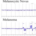 Cytogenetic and Mutational Analyses of Melanocytic Tumors
 Dermatology Clinics
 Update on Vascular Neoplasms
Current Understanding of Cutaneous Lymphoma
Direct Immunofluorescence Testing in the Diagnosis of Immunobullous Disease, Collagen Vascular Disease, and Vascular Injury Syndromes
New Directions in Dermatopathology
Update on Vascular Neoplasms
Current Understanding of Cutaneous Lymphoma
Direct Immunofluorescence Testing in the Diagnosis of Immunobullous Disease, Collagen Vascular Disease, and Vascular Injury Syndromes
New Directions in Dermatopathology
Stay updated, free articles. Join our Telegram channel

Full access? Get Clinical Tree


