In vivo confocal microscopy represents a new device that generates a virtual skin biopsy at cytologic resolution. This article describes the most relevant confocal findings and their histopathologic correlates in skin oncology and inflammatory diseases. The light and dark of confocal microscopy are briefly discussed in relation with its clinical applications.
- •
Reflectance confocal microscopy (RCM) is a novel noninvasive imaging technique that enables the identification of cells and tissues with nearly histologic resolution.
- •
In the epidermis, keratinocytes are clearly visualized as small polygonal nucleated structures, and epidermal alterations, such as acanthosis, hyperkeratosis, exocytosis, and spongiosis, and others can be easily identified.
- •
RCM also enables the visualization of dermal structures up to the papillary dermis.
- •
In melanocytic and epithelial skin tumors, cytologic atypia as well as architectural disarrangement can be visualized, helping in the achievement of a more accurate diagnosis.
- •
The application in inflammatory and infectious skin diseases showed good correlation with microscopic findings, although inflammatory cell subpopulations cannot be distinguished.
Introduction
Reflectance confocal microscopy (RCM) represents a new imaging tool that enables the identification of cells and tissues with nearly histologic resolution. Although several noninvasive tools have been explored to test their potential application in the clinical field, RCM has emerged as a unique instrument because it can visualize the skin tissue with a resolution that is comparable with conventional histopathology. It allows a horizontal scanning of the imaged tissue, with the advantage of exploring a larger field of view compared with vertical sectioning. Moreover, the horizontal plane offers a perfect correlation with clinical and dermoscopic aspects, which is crucial when dealing with skin tumor diagnosis. In this article, we present the main confocal findings and their correlations with histopathology along with a brief description of confocal applications in the clinic arena.
Instruments
The commercially available confocal microscope (VivaScope 1500, Lucid, Rochester, NY) contains a probe (the head of the microscope), which is attached to the skin by using a disposable plastic window, which is in turn taped to a metal ring. A confocal microscope consists of a point source of light, condenser, objective lenses, and a point detector. The pinhole collects light emanating only from the in focus plane. The mechanism of bright contrast in RCM is backscattering. In gray-scale confocal images, structures that appear bright (white) have components with high refractive index compared with their surroundings and are similar in size to the wavelength of light. Backscattering is primarily governed by the refractive index of the structure compared with surrounding medium. Highly reflective skin components include melanin, collagen, and keratin. The confocal scanning produces high-resolution black-and-white horizontal images (0.5 × 0.5 mm) with a lateral resolution of 1.0 μm and axial resolution of 3 to 5 μm. A sequence of full-resolution individual images at a given depth is acquired and stitched together to create a mosaic ranging in size from 2 × 2 mm to 8 × 8 mm. For inflammatory or physiologic skin conditions, a 3 × 3 mm VivaCube (Lucid, Rochester, NY) composed of 4 mosaics with a 25-μm step is usually acquired, whereas for skin tumor examination, the imaging should include the entire lesion. Besides the horizontal mosaic, a vertical VivaStack can be imaged. It consists of single high-resolution images acquired from the top skin surface up to 200 μm, corresponding to the papillary dermis, to obtain a sort of optic biopsy. The VivaStack (Lucid, Rochester, NY) modality is useful for the assessment of the epidermal thickness either in physiologic condition or in the presence of dysfunctional epidermis.
Recently, a handheld RCM has been introduced on the market (VivaScope 3000). This version is a smaller, flexible device, which is useful in areas that are difficult to access (eg, skin folds, ears). Unlike the 1500 version, it has an on-instrument control for laser power, imaging depth, and capture but it does not allow scanning of a large field of view, which is needed, for example, in some tumors to obtain an overview of the architecture. However, it is a promising tool, which can be used for surgical premapping or when multiple site imaging is requested.
Main histopathologic correlates of confocal criteria
Epidermis
The epidermis can be affected by several injuries that lead to different morphologic changes involving the keratinocytes (KCs) or other cells of the epidermis, such as melanocytes.
The epidermal changes are described as phenomenon per se regardless of their relationship with either inflammatory or skin tumors.
In healthy young skin, the epidermis appears as a multilayer tissue with paradigmatic confocal aspects depending on the skin level. The stratum corneum appears as a highly refractive surface surrounded by visible skin furrows. At this level, the corneocytes are large, ranging from 10 to 30 μm, polygonal, and without a visible nucleus. Skin furrows appear as dark folds between islands of KCs. In young people, the skin furrows are arranged in a rhomboidal pattern formed by intersecting skin furrows. However, the shape and arrangement of the skin folds strongly depends on the body site (being almost absent on the forehead and well represented on the abdomen) and the individual’s age. The stratum granulosum is composed of polygonal KCs presenting a bright and grainy cytoplasm because of the presence of organelles. The KCs cohesively assemble, forming a structure that gives rise to a honeycombed pattern. The contour of the cell is usually brighter than the cytoplasm and perfectly outlined. Pigmented KCs are usually bright cells, small in size and always polygonal, separated by a darker contour (cobblestone pattern), resembling the negative of the honeycombed pattern. In the honeycombed pattern, the KCs appear black with bright contour, whereas in the cobblestone pattern, these cells show a bright cytoplasm because of the high melanin content (ie, brightness) ( Fig. 1 ). On the face, a peculiar pattern is caused by the presence of numerous hair follicles that appear as dark round areas (donutlike appearance, H Rabinovitz, personal observation). At the stratum spinosum level, the size of KCs tends to decrease but the cells still have a polygonal shape. The honeycombed pattern is easily observable.
Acanthosis is one of the most frequent findings and it can be observed in several conditions. Histopathologically, acanthosis is defined as diffuse epidermal hyperplasia caused by the increased thickness of the stratum spinosum constituted by the prickle cells.
Because of the horizontal sectioning, only an indirect correlate with acanthosis is feasible on RCM. The granulosum and spinosum layers of an acanthotic epidermis consist of islands of KCs with broadened greyish outlines ( Fig. 2 ). Moreover, when using the VivaStack modality, an increased thickness of the stratum granulosum/spinosum can be detected and measured by counting the layers.
Dermoepidermal Junction
Below the spinous layer, there is a single layer of basal cells at the dermoepidermal junction (DEJ). Basal cells are uniform in size and shape but are smaller and more refractive than spinous KCs because of the melanin caps forming bright disks on top of the nuclei. The brightness of KCs is strongly dependent on the skin phototype. Dark skin phototypes show basal KCs that are bright round cells with highly refractive cytoplasm forming a cobblestone pattern at the epidermal level and bright rings at the dermoepidermal junction level. On the other hand, skin phototype I-II are characterized by basal KCs with low refractivity that constitute barely visible dermal papillae ( Fig. 3 ). At the dermoepidermal level, in the presence of regular rete-ridges, basal cells form round or oval rings of bright cells (KCs) surrounding dark dermal papillae. Within the dark dermal papillae, it is possible to visualize tiny canalicular blood vessels.
In solar lentigos and in seborrheic keratosis, the DEJ reveals small bud-acanthotic proliferations, often anastomosing, and hyperpigmentation of the basal layer. This histologic finding translates into polycyclic bright contours or bulbous projections caused by the anastomosing elongated structures, separated by dark areas ( Fig. 4 ), on horizontal plane RCM.
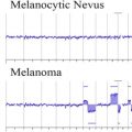
In skin cancer and in melanocytic proliferations, the DEJ is often distorted and occupied by tumor proliferations. In nevi, junctional nests can be seen as compact, well-outlined, roundish-to-oval structures, sometimes connected with the epidermis. In some instances, a few atypical large and bright cells can occasionally be found in nevi. Many junctional nests form the so-called meshwork pattern ( Fig. 5 ). This pattern is made by the enlargement of the interpapillary spaces formed by aggregated cells or clusters bulging within the dermal papilla in contiguity with the basal layer. When several nests are located within the dermal papillae, the general architecture is called clod pattern and corresponds to the presence of dermal nests seen in the superficial dermis in compound nevi ( Fig. 6 ). In malignant melanoma, the DEJ can show areas of flattening and disarrangement of the architecture, responsible for fuzzy and not well-defined papillary contours (so-called nonedged papillae), alternating with single-cell proliferations or nests of large pleomorphic atypical cells (aspecific pattern, Fig. 7 ).
Papillary Dermis
In the papillary dermis, the main findings that can be evaluated with high resolution using RCM are the inflammatory infiltrate, the regression phenomenon, and solar elastosis. The tumoral proliferation of the dermal compartment is reported in a specific section.
The inflammatory infiltrate is constituted by distinct cell types. On RCM, we can readily detect melanophages and lymphocytes. Melanophages appear as plump, large, bright cells with ill-defined borders and no visible nucleus. These cells are usually seen within the dermal papillae or in the dermis and are often clustered ( Fig. 8 ). In the regression phenomenon, melanophages are seen admixed with coarse and bright collagen fibers corresponding to a fibrotic response. Occasionally, a few melanocytes can be found, but their distinction from the inflammatory infiltrate is not always feasible in regressive areas ( Fig. 9 ). Lymphocytes show up as small bright triangular structures, which can be focal or widespread. In aged skin or heavily sun-damaged skin, it is possible to detect the presence of curled bright fibers that correspond to fragmented elastic fibers on histochemistry.
Main clinical applications of in vivo confocal microscopy
Skin Tumors
In skin oncology, the goal is to make an early diagnosis and reduce the number of unnecessary biopsies. Because the skin is easy to access, several instruments have been applied in oncology, although the gold standard in clinical practice is considered the combination of clinical inspection and dermoscopy. Dermoscopy is a noninvasive and cheap technique, which has proved to be an essential tool in skin oncology and general dermatology. RCM represents a second-level examination in clinical practice, which found its application in challenging cases or in special instances. In addition to dermoscopy, RCM is capable of delivering single-cell resolution in a few minutes at the patient’s bedside.
Melanocytic Tumors
In common nevi, RCM reveals a symmetric architecture that is characterized by junctional nests (meshwork pattern, see Fig. 5 ) or dermal nests (clod pattern, see Fig. 6 ) with little or no cytologic atypia. When dealing with the so-called gray zone constituted by atypical or dysplastic nevi, a puzzling picture is shared by both RCM and histopathology. On RCM, it is possible to detect some morphologic aspects such as bridging of the nests or the presence of atypical cells, but it is not always feasible to draw a line between a severe dysplastic nevus and an incipient melanoma. This challenge relies on the clinician’s ability to read the confocal image or on their belief in the existence of a precursor lesion. The same scenario is present also in dermatopathology and accounts for the low interobserver agreement found when analyzing these kinds of pigmented lesions. Spitzoid lesions represent another challenging situation in clinical practice. Although the confocal features of Spitz nevi are partially characteristic, they remain in most cases indistinguishable from melanoma. A good correlation was found for some histopathologic aspects and RCM features, some of which are considered characteristic of Spitz nevi, such as sharp lateral demarcation and presence of spindled cells. Other correlates that are not specific include pagetoid infiltration, junctional and dermal nests, parakeratosis, transepidermal melanin elimination, and inflammatory infiltrate rich in melanophages. However, no correlate was found for other characteristic histologic aspects, such as Kamino bodies and maturation with depth. The frequent presence of features suggestive of malignancy in Spitz nevi, such as pagetoid infiltration ( Fig. 10 ) and atypical cells, nonedged papillae, dishomogeneous nests, and the impossibility to explore the deeper parts of a given lesion, hamper a reliable diagnosis with RCM.
Related posts:
 Cytogenetic and Mutational Analyses of Melanocytic Tumors
 Dermatology Clinics
Fibrous and Fibrohistiocytic Neoplasms
Histopathologic Patterns Associated with External Agents
Current Understanding of Cutaneous Lymphoma
Direct Immunofluorescence Testing in the Diagnosis of Immunobullous Disease, Collagen Vascular Disease, and Vascular Injury Syndromes
Dermatology Clinics
Fibrous and Fibrohistiocytic Neoplasms
Histopathologic Patterns Associated with External Agents
Current Understanding of Cutaneous Lymphoma
Direct Immunofluorescence Testing in the Diagnosis of Immunobullous Disease, Collagen Vascular Disease, and Vascular Injury Syndromes
Stay updated, free articles. Join our Telegram channel

Full access? Get Clinical Tree


