Fig. 65.1
ENTL, nasal type: Ulcerated tumor in the nasal area (Courtesy of Dr. Alistair Robson, London, UK)
Pathology
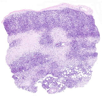
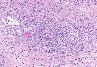
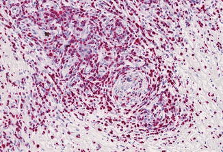
ENTL is characterized by dense dermal and subcutaneous infiltrates with an angiocentric and angiodestructive growth resulting in necroses and ulceration (Figs. 65.2 and 65.3). The tumor cells are of variable size ranging from small to large cells with pleomorphic nuclei and pale cytoplasm (Fig. 65.3). Mitoses are common. In some cases, numerous admixed reactive cells such as eosinophils, plasma cells, and histiocytes are observed. The tumor cells express CD2, cytoplasmic CD3 (CD3e), CD56, as well as cytotoxic proteins (T1A-1, granzyme B, and perforin). Presence of EBV in the neoplastic cells can be demonstrated by in situ hybridization in nearly all cases of secondary cutaneous NK/T- and NK-cell lymphomas (Fig. 65.4) but is reported to be rarely found in primary cutaneous forms of ENTL. Most cases are in germline configuration, but clonal T-cell receptor gene rearrangement can be detected in a minority of the cases.


Fig. 65.2
ENTL, nasal type: Dermal and subcutaneous infiltrates

Fig. 65.3
ENTL, nasal type: Angiocentric and angiodestructive infiltrates of medium-sized to large tumor cells with pleomorphic nuclei

Fig. 65.4
ENTL, nasal type: Detection of EBV in all tumor cells by in situ hybridization
Differential Diagnosis
Differential diagnosis includes other CD56+ and/or EBV-associated T- or NK-cell lymphomas as well as lymphomas with angioinvasive growth pattern including cutaneous gamma/delta T-cell lymphoma, pagetoid reticulosis, subcutaneous panniculitis-like T-cell lymphoma, and primary cutaneous or secondary cutaneous peripheral T-cell lymphoma, unspecified. Immunophenotyping and especially detection of EBV are useful to distinguish ENTL from the abovementioned lymphomas. EBV-associated mucocutaneous ulcer is a B-cell lymphoproliferation often arising in the context of drug-related immunosuppression which differs by the morphology and B-cell phenotype of the atypical lymphoid cells resembling Reed-Sternberg or Hodgkin-like cells and the excellent prognosis after withdrawal of underlying immunosuppression.
Stay updated, free articles. Join our Telegram channel

Full access? Get Clinical Tree


