Fig. 16.1
Slowly enlarging, well demarcated, erythematous, eczematous plaque in the left inguinal area and on the scrotum
Pathology
EMPD is an asymmetrical, multicentric, poorly demarcated neoplasm in which the atypical epithelial cells are disposed within the epidermis and epithelial structures of adnexa. The cells are arranged as solitary units and in nests of different sizes in the basal layer, throughout the epidermis, and within the depth of epithelial structures of eccrine ducts and folliculosebaceous units (Fig. 16.2). Nests of Paget cells are not equidistant from one another and have a tendency to confluence and to form glandular structures. Most cells are concentrated in the lower portion of the epidermis and in the epithelial structures of adnexa. The neoplastic cells have round or oval pleomorphic nuclei, prominent nucleoli, and abundant pale cytoplasm with a bluish cast due to intracytoplasmic mucin. This may not always be apparent on routine sections. Occasionally, Paget’s cells can take on a signet-ring appearance due to intracytoplasmic accumulation of mucin (Fig. 16.3). Signs of apocrine differentiation in the form of glandular formation with decapitation secretion and mucinous cells are sometimes seen (Fig. 16.4). An underlying adnexal adenocarcinoma may occasionally be found.
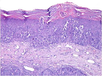
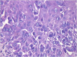
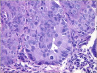
Fig. 16.2
The atypical epithelial cells are disposed as solitary units and in nests throughout the epidermis, mostly in the lower portion of it
Fig. 16.3
The neoplastic cells have round or oval pleomorphic nuclei, prominent nucleoli, and abundant pale cytoplasm with a bluish cast. Occasionally, the cells take on a signet-ring appearance due to intracytoplasmic accumulation of mucin
Fig. 16.4
Glandular formation with hints of decapitation secretion is also seen. Many mitotic figures, including atypical forms, are easily identified
Histochemistry
Paget cells are diastase-resistant, PAS-positive, Alcian blue-positive, mucicarmine-positive, aldehyde fuchsin-positive, and toluidine blue-positive cells. Apocrine differentiation markers α-1-antitrypsin and lysozyme are commonly expressed by the tumor cells.
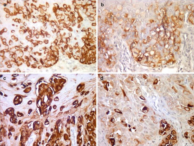
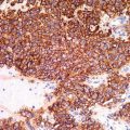
Immunohistochemistry
The tumor cells of EMPD usually express CK5/6, CK7, CEA, EMA, AR, Her2neu, and GCDFP-15 (Fig. 16.5a–d) and are negative for ER, PR, bcl-2, and c-erbB-2. CK20 is usually negative in primary cutaneous EMPD and positive in secondary EMPD. CDX2 for colon, PSA for prostate, and uroplakin for bladder carcinoma are useful markers in the evaluation of EMPD with an underlying tumor.