The eyes and periocular area are the central aesthetic unit of the face. Facial aging is a dynamic process that involves skin, subcutaneous soft tissues, and bony structures. An understanding of what is perceived as youthful and beautiful is critical for success. Knowledge of the functional aspects of the eyelid and periocular area can identify pre-preoperative red flags.
Key points
- •
The eyes and periocular area are the central aesthetic unit of the face.
- •
Facial aging is a dynamic process that involves skin, subcutaneous soft tissues, and bony structures.
- •
An understanding of what is perceived as youthful and beautiful is critical for success.
- •
Knowledge of the functional aspects of the eyelid and periocular area can identify preoperative red flags.
Introduction
The beauty of a woman must be seen from in her eyes, because that is the doorway to her heart, the place where love resides
Appearance has an important role in self-perception as well as perception by others. No area is more important in self-perception than the face, which research has shown to have a profound effect on overall well-being. As the center of the face, the functional and aesthetic importance of the periocular region cannot be overstated. Not only is the periocular area the core aesthetic unit of the face, it is also responsible for protection and function of the eye and thus the visual system. Periorbital aging changes are among the earliest to present in the face, which can cause patients distress with even small changes to this area.
Patient attention to the periorbital region has driven cosmetic blepharoplasty to become one of most commonly performed surgical procedures in the world. Although complication rates with periorbital aesthetic treatments are generally low, visually threatening events can occur. Given the intricate relationship of the periorbital area with the visual system, an intimate understanding of anatomy combined with a thorough preoperative evaluation and meticulous surgical technique are essential to provide the highest patient outcomes. The preoperative evaluation is crucial to assessing patient goals, establishing the surgical plan, setting realistic expectations, and identifying any risk factors that could lead to poor outcomes. The following is our standard approach to evaluating the functional and aesthetic issues in the periorbital area.
The beautiful eye
Symmetry
Symmetry is of paramount importance in the perception of facial beauty. Humans have a sensitive perception of symmetry, the ability to detect perfect symmetry, and to discern very small amounts of asymmetry. The correlation between symmetry and attractiveness is significant, with studies finding digitalized mirror-image faces perceived as more attractive than the unaltered original images. Thus, the importance of symmetry to oculofacial plastic surgeons cannot be overstated.
The periocular region frequently has visible asymmetry in the brow, eyelid height, amount of skin redundancy, cheek projection, and globe prominence. Although there is no general consensus on the amount of asymmetry that is clinically significant, previous studies have found significant asymmetry in up to 30% of patients. Many patients are unaware of their asymmetries, which leads to poor surgical outcomes if not addressed properly addressed at surgery. Surgeons must review with their patients, document all asymmetry, and factor its role into the treatment plan. Patients must be counseled that at least a small degree of asymmetry will persist postoperatively. Unrealistic expectations can lead to marked patient dissatisfaction.
Aging
Ideally, balanced and diffuse fat distribution, a well-rounded three-dimensional structure, and good projection are hallmarks of a healthy and youthful facial appearance. Little described the youthful face as an ogee-shaped profile, with anterior oblique surfaces that undulate in graceful curves.
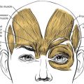
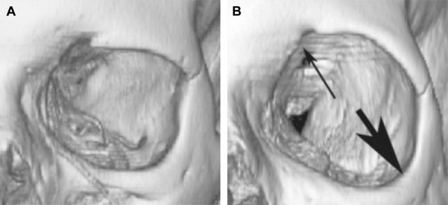
Facial aging is a dynamic process that involves skin, subcutaneous soft tissues, and bony structures. The bony remodeling of the orbit results in orbital elongation, loss of projection, and change of the bone–soft tissue relationship ( Fig. 1 ), which likely contributes to the fat prolapse, hollow sulci, ptosis, brow descent, and lateral upper eyelid hooding that is commonly seen in aging. The skin and subcutaneous tissues around the periocular area become increasingly hollow, allowing the underlying bone, muscle, remaining fat, and blood vessels to become more apparent. The gradual descent and atrophy of subcutaneous tissue alters the smooth ogee curve of the youthful face.
Older surgical techniques for periocular rejuvenation are generally purely subtractive, with the removal of fat and skin, which led to an increasing hollow appearance. However, the increased understanding of the aging process has resulted in the amelioration and development of new techniques leading to more successful rejuvenation outcomes.
Skin
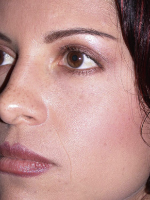
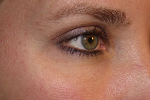
Skin is the largest organ in the human body. Healthy skin tone, a smooth appearance, and brighter complexion are associated with increased attractiveness, youth, and health. Because wrinkles are a sign of aging, youthful skin should be smooth. Ideally, the pigment is uniform and skin is free of blemishes with a consistent appearance ( Fig. 2 ).
Upper eyelid
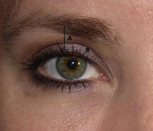
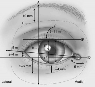
The upper eyelid margin naturally has a gently curved contour. Medially, the curve has a sharper angle with the peak height located between the pupil and lateral limbus in the Western eyelid ( Fig 3 ). The central height of the upper eyelid should be just below the limbus, without excessive droop or scleral show. The ideal upper eyelid should not have excess fat, or skeletonization with hollowing of the sulcus ( Fig. 4 ). The youthful aesthetics of the Asian upper eyelid are different in numerous ways, including the eyelid fold, lower marginal position, and more temporal peak in the lid height.
Upper Eyelid Height
Described by Putterman and Urist in 1975, the margin reflex distance (MRD) is a widely used and specific way to evaluate upper eyelid position. MRD can replace palpebral fissure because palpebral fissure does not examine the upper and lower eyelid heights independently. To determine the MRD, the surgeon shines a light held at the surgeon’s eye level toward the patient’s eyes in primary gaze, looking at the corneal light reflex. The MRD1 is the measurement from the corneal light reflex to the central upper eyelid margin. The MRD2 is similarly the measurement from the corneal reflex to the central lower eyelid margin. To ensure accurate measurement, surgeons must ensure that the patient’s brow is in a natural position and that the surgeon’s and patient’s eyes are at the same level.
Although there is variability between ethnic groups, the normal Western MRD1 is 3.5 mm to 4.5 mm. Any asymmetry between the 2 eyes, or an abnormal MRD1, must be addressed. Many patients are unaware preoperatively and a blepharoplasty in the setting of blepharoptosis makes the ptotic eyelid position more apparent.
Levator Function
Levator function is the measurement of upper eyelid excursion from downgaze to upgaze. Ensuring that eyebrow excursion does not contribute to the levator function is key to an accurate measurement and is accomplished by manually holding the eyebrow in its normal position. Normal levator function is greater than 12 mm, with an average of 15 mm. A significant decrease in levator function can indicate congenital or myopathic ptosis.
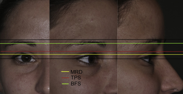
Upper Eyelid Crease, Tarsal Platform Show, and Brow Fat Span
A normal, properly defined eyelid crease for white people is 10 to 12 mm above the central eyelid margin in women, and 7 to 8 mm in men. There are notable variations among different ethnic groups.
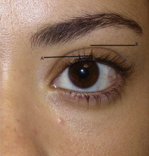
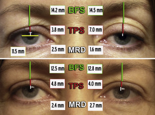
Tarsal platform show (TPS) is the amount of fixed pretarsal skin that is visible inferior to the skin overlying the eyelid crease, which is normally 3 to 6 mm in Western Europeans. Brow fat span is the distance between the skin fold overlying the eyelid crease (top edge of the TPS) to the inferior brow hairs ( Fig. 5 ). Attention must be paid to the symmetry of these measurements ( Fig. 6 ), because TPS symmetry may be more important than MRD1 in eyelid symmetry perception.
Lower eyelid
The lower eyelid spans from the lateral to medial canthus. The lateral canthal angle is slightly higher than the medial canthus ( Fig. 7 ). The lower eyelid continues with a gentle curve with the ideal position of the central margin at or slightly above the inferior limbus. The thin eyelid skin transitions seamlessly to the thicker cheek inferiorly. A small area of pretarsal orbicularis muscle just inferior to the lashes gives a healthy, youthful appearance.
The transition between the eyelid and cheek, commonly referred to as the nasojugal fold or tear trough, should be a smooth, gradual transition without a hollow deformity ( Fig. 8 ). Although complicated and multifactorial, a combination of facial volume loss, herniation of orbital fat, inferolateral rim remodeling, skin laxity, and ptosis of midface tissues contributes to the common tear trough deformity seen in the aging lower eyelid.