Erythroderma


Abstract


From a diagnostic and therapeutic perspective, erythroderma represents one of the most challenging entities within dermatology. This is due in part to the large number of disorders that can present as an erythroderma. Whilst in adults the commonest causes are inflammatory disorders such as atopic dermatitis and psoriasis, drug eruption should always be in the differential diagnosis. In children, a variety of rare genetic disorders also need to be considered. Careful clinicopathologic correlation is essential in order to identify specific underlying causes and extensive investigations are often required to exclude disorders such as cutaneous T-cell lymphoma. Despite a thorough evaluation, a significant minority of patients (~25%) with erythroderma may have no identifiable etiology. While treatment should be directed at the underlying cause, weekly methotrexate can be an excellent empiric therapeutic option for patients with idiopathic erythroderma.


Keywords

erythroderma, exfoliative dermatitis, exfoliative erythroderma, drug eruption, drug reaction, Sézary syndrome, atopic dermatitis, psoriasis, idiopathic erythroderma, erythrodermic mycosis fungoides, pityriasis rubra pilaris


Synonyms


▪ Exfoliative dermatitis ▪ Exfoliative erythroderma ▪ Red man syndrome


Key features


  • Erythroderma is clinically defined as erythema and scaling involving >80–90% of the body surface area


  • Systemic manifestations include peripheral edema, tachycardia, loss of fluid and proteins, and disturbances in thermoregulation


  • Erythroderma has multiple etiologies; the most common causes are psoriasis, drug reactions, atopic dermatitis, and cutaneous T-cell lymphoma (CTCL)


  • Establishing the correct diagnosis requires consideration of initial sites of involvement, additional clinical findings, histologic and molecular features, and associated systemic abnormalities, as well as a complete medical history


  • Despite an intensive evaluation, the cause remains unknown (idiopathic) in 25–30% of patients; some of these patients eventually develop CTCL


  • Treatment strategies should address the dermatologic disease as well as the underlying etiology and the systemic complications of the erythroderma


Introduction


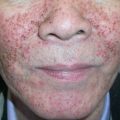
Erythroderma is defined as generalized erythema and scaling involving >80–90% of the body surface area (BSA). However, it does not represent a defined entity, but rather is a striking clinical presentation that can arise from a variety of diseases. Most commonly, erythroderma is due to generalization of pre-existing dermatoses (such as psoriasis or atopic dermatitis), drug reactions, or cutaneous T-cell lymphoma (CTCL). Although up to 50% of the patients have a history of more localized skin lesions prior to the onset of the erythroderma, identification of the underlying disease process represents one of the most complex challenges in dermatology. Sustained efforts during longitudinal evaluation may lead to the precise identification of the etiology. In approximately one-quarter of the patients, no specific etiology is found, and these cases are called “idiopathic erythroderma”.


Attention should also be focused on the potential systemic complications of acute erythroderma. Hypothermia, peripheral edema, and loss of fluid, electrolytes and albumin with subsequent tachycardia and cardiac failure are serious threats to the erythrodermic patient. In addition, chronic erythrodermas may be accompanied by cachexia, diffuse alopecia, palmoplantar keratoderma, nail dystrophy, and ectropion.


Historical Perspective


The term “erythroderma” was introduced in 1868 by Hebra to describe an exfoliative dermatitis involving more than 90% of the skin surface. Based upon the clinical course, erythroderma was classified into chronically relapsing (Wilson–Brocq), chronically persisting (Hebra), and self-limiting epidemic (Savill) variants. However, these subdivisions are no longer employed. Even though originally more strictly defined as erythema and scaling involving >90% of BSA, nowadays the term erythroderma is often more liberally applied when there is >80% of BSA involvement.


Epidemiology


No precise data exist regarding the prevalence or incidence of erythroderma as most reports are retrospective and do not address the issue of overall incidence. Large series of patients have focused on male-to-female ratios, average age, and underlying diseases . Men are more commonly affected, with the male-to-female ratio ranging from approximately 2 : 1 to 4 : 1. An even higher ratio can be found in the subset of idiopathic erythroderma, also referred to as “red man syndrome” (not to be confused with the acute cutaneous reaction to rapid infusion of vancomycin). The average age at onset of erythroderma in these series was 52 years, with an average of 48 years in those including children, and 60 years in series excluding them .


Of a total of 746 patients, dermatitis (24%), psoriasis (20%), drug reactions (19%), and CTCL (8%) represented the most common underlying causes of erythroderma . When categories within the dermatitis group were examined, atopic dermatitis (9%) was the most common type, followed by contact dermatitis (6%), seborrheic dermatitis (4%), and chronic actinic dermatitis (3%). With regard to etiology, no specific geographic differences have been noted . In adults with erythroderma, overall relapse rates at one year range from 20% to 30%.


For adults, uncommon to rare causes include pityriasis rubra pilaris, ichthyoses, bullous dermatoses (usually pemphigus foliaceus), graft-versus-host disease (GVHD), infestations (most often scabies), and autoimmune connective tissue diseases (acute or subacute lupus erythematosus, dermatomyositis). Table 10.1 lists additional rare causes, from paraneoplastic (e.g. lymphoma) to inflammatory (e.g. sarcoidosis) and neoplastic (e.g. mastocytosis). Despite multiple skin biopsies, an in-depth clinical investigation and a detailed medical history, the underlying cause of erythroderma is not found in at least 25% of patients. Unfortunately, cases of idiopathic erythroderma tend to be chronic and are more likely to recur after treatment .


Table 10.1

Causes of erythroderma in adults.

BMZ, basement membrane zone; DDI, dideoxyinosine; DIF, direct immunofluorescence; DIHS, drug-induced hypersensitivity syndrome; DRESS, drug reaction with eosinophilia and systemic symptoms; GVHD, graft-versus-host disease; HIV, human immunodeficiency virus; IIF, indirect immunofluorescence.


CAUSES OF ERYTHRODERMA IN ADULTS
Underlying disease Clinical clues Histologic clues Additional hints
Common
Psoriasis ( Ch. 8 )


  • Pre-existing psoriatic plaques or lesions of pustular psoriasis


  • Often spares the central face


  • Nail changes (oil-drop, pits, onycholysis)


  • Subcorneal pustules


  • Inflammatory arthritis


  • Confluent or focal parakeratosis


  • Epidermal hyperplasia with bottleneck-like rete ridges


  • Tortuous vessels in a slightly edematous papillary dermis


  • Neutrophils within epidermis; sparse lymphohistiocytic infiltrate within dermis


  • Often reduced or absent granular layer


  • Personal or family history of psoriasis


  • Withdrawal of corticosteroids, methotrexate, cyclosporine or targeted immune modulators (biologic agents)


  • Receiving medication that can exacerbate psoriasis (e.g. lithium)

Atopic dermatitis ( Ch. 12 )


  • Pre-existing lesions (flexures)


  • Severe pruritus


  • Lichenification, including eyelids


  • Prurigo nodularis


  • Mild to moderate acanthosis


  • Variable spongiosis


  • Dermal eosinophils


  • Parakeratosis


  • Variable lymphohistiocytic infiltrate with exocytosis of lymphocytes


  • Variable dermal edema


  • Elevated serum IgE, eosinophilia


  • Personal or family history of atopy (e.g. asthma, allergic rhinitis, atopic dermatitis)


  • Cataracts/keratoconus

Drug reactions ( Ch. 21 )


  • Preceded by morbilliform or scarlatiniform exanthem


  • Facial edema


  • In dependent areas, may become purpuric


  • Perivascular infiltrate with eosinophils


  • Lichenoid pattern


  • Lymphomatoid pattern


  • Spongiotic pattern


  • Apoptotic keratinocytes at all levels of epidermis


  • Vacuolar degeneration of basal layer


  • DRESS/DIHS – spongiotic and lichenoid patterns


  • No history of skin diseases


  • More acute onset


  • Usually resolves within 2–6 weeks after withdrawal of responsible drug; possible exception is DRESS/DIHS


  • More common in HIV-infected patients

Idiopathic erythroderma


  • Elderly men


  • Chronic, relapsing


  • Severe pruritus


  • Palmoplantar keratoderma


  • Dermatopathic lymphadenopathy


  • Nonspecific


  • Acute forms – parakeratosis, spongiosis, and a moderate lymphohistiocytic infiltrate


  • Chronic forms – hyperkeratosis, psoriasiform acanthosis, and expanded papillary dermis


  • Consider less commonly associated drugs (see Table 10.3 )


  • Continue to re-evaluate for cutaneous T-cell lymphoma, including serial skin biopsies

Less common
Cutaneous T-cell lymphoma (Sézary syndrome > erythrodermic mycosis fungoides; Ch. 120 )


  • Intense pruritus with excoriations; can develop lichenification or prurigo nodularis


  • Deep purple–red hue; pigmentary changes (melanoerythroderma)


  • Infiltration and/or edema; leonine facies


  • Painful, fissured palmoplantar keratoderma


  • Alopecia


  • Lymphadenopathy


  • Cerebriform pleomorphic lymphocytes


  • Clustering of atypical cells within epidermis uncommon


  • Band-like and occasionally lichenoid infiltrate


  • Cutaneous histology is non-diagnostic in at least 30% of patients with Sézary syndrome


  • Peripheral blood CD4 + lymphocytosis in Sézary syndrome (elevated CD4 : CD8 ratio of ≥10 : 1 in blood)


  • Detection of identical clonal T-cell population in skin and lymph node/blood


  • In Sézary syndrome: ≥1000 Sézary cells/mm 3 ; ≥40% CD4 + /CD7 ; or ≥30% CD4 + /CD26 (blood)


  • Can mimic several other causes of erythroderma

Pityriasis rubra pilaris ( Ch. 9 )


  • Salmon-colored erythema


  • Islands of sparing (nappes claires)


  • Waxy keratoderma


  • Perifollicular keratotic papules


  • Psoriasiform epidermal hyperplasia


  • Granular layer is normal


  • Alternating parakeratosis and orthokeratosis, both vertically and horizontally


  • Follicular plugs with “shoulder parakeratosis”


  • No neutrophils


  • Flare after sun exposure


  • Cephalocaudal progression


  • Appearance of erythema gyratum-like lesions as erythroderma improves

Dermatitis (non-atopic), including contact ( Chs 14 & 15 ) and stasis with autosensitization ( Ch. 13 )


  • Pre-existing localized disease


  • Distribution of initial lesions


  • Variable spongiosis


  • Occupation and hobbies


  • Patch testing


  • Review oral medications (systemic contact dermatitis)

Paraneoplastic erythroderma


  • Fine scaling


  • Melanoerythroderma


  • Cachexia


  • Nonspecific changes


  • Lymphoproliferative disorders, including lymphomas other than Sézary syndrome, and rarely thymomas


  • In the case of solid-organ malignancies, usually late-stage

Bullous dermatoses ( Chs 29 & 30 ) and inherited ichthyoses ( Ch 57 )
Pemphigus foliaceus


  • Pre-existing lesions, often on upper trunk


  • Impetigo-like erosions and vesicles


  • Moist scale-crust; cornflake scale


  • Acantholysis within the superficial epidermis


  • DIF, intercellular IgG


  • IIF, intercellular

Bullous pemphigoid


  • Urticarial plaques


  • Tense bullae


  • Elderly patients


  • Subepidermal blister


  • Eosinophils


  • DIF, BMZ C3 and IgG


  • IIF, BMZ

Paraneoplastic pemphigus


  • Mucosal erosions and hemorrhagic crusts


  • Erythema multiforme-like lesions


  • Mucocutaneous lichenoid lesions


  • Interface dermatitis with necrotic keratinocytes


  • Parakeratosis


  • Focal acantholysis


  • DIF, intercellular and BMZ IgG


  • IIF, intercellular and BMZ (+ IIF rat bladder)

Inherited ichthyoses ( Ch. 57 )


  • Present from birth or early infancy (see Table 10.2 )


Rare
Papuloerythroderma of Ofuji


  • Widespread, pruritic, flat-topped, red–brown papules that can become confluent


  • Sparing of skin folds (“deck-chair” sign)


  • Favors elderly men


  • Dense perivascular infiltrate of lymphocytes and eosinophils in the upper to mid dermis


  • Multiple etiologies, including CTCL > atopy, other lymphomas, drugs (e.g. DDI, aspirin), infections (e.g. HIV, hepatitis C virus), paraneoplastic phenomenon


  • May have peripheral eosinophilia, lymphopenia

Chronic actinic dermatitis ( Ch. 87 )


  • Initial lesions in photodistribution


  • Allergic airborne contact dermatitis to Compositae


  • Lichenoid infiltrate of lymphocytes and epidermal exocytosis


  • Acanthosis of epidermis and compact hyperkeratosis


  • Possible lymphocyte nuclear pleomorphism


  • Drug history


  • UVA, UVB and visible light phototesting


  • Photopatch testing

Other rare causes


  • Hypereosinophilic syndrome ( Ch. 25 )


  • Crusted (Norwegian) scabies ( Ch. 84 )


  • Lichen planus


  • GVHD ( Ch. 52 )


  • Autoimmune connective tissue disease (e.g. acute or subacute cutaneous lupus erythematosus, juvenile dermatomyositis [adolescents])


  • Dermatophyte infection


  • Primary immunodeficiencies ( Ch. 60 )


  • Sarcoidosis


  • Mastocytosis


  • Langerhans cell histiocytosis


  • Other T-cell hematologic malignancies (e.g. adult T-cell leukemia/lymphoma, angioimmunoblastic T-cell lymphoma, T-cell prolymphocytic leukemia)


With regard to neonates and infants, inherited ichthyoses, dermatitides, psoriasis, immunodeficiencies (e.g. Omenn syndrome), and consequences of infection (e.g. staphylococcal scalded skin syndrome) represent the major causes of erythroderma ( Table 10.2 ) . In addition, the possibility of drug-induced erythroderma should always be considered.


Table 10.2

Causes of erythroderma in neonates and infants.

ELISA, enzyme-linked immunosorbent assay; IPEX, immune dysregulation, polyendocrinopathy, enteropathy, X-linked; MRSA, methicillin-resistant Staphylococcus aureus ; SCID, severe combined immunodeficiency; TSST-1, toxic shock syndrome toxin 1.


CAUSES OF ERYTHRODERMA IN NEONATES AND INFANTS
Underlying disease Clinical clues Histologic clues Additional findings
Inherited ichthyoses ( Ch. 57 )
Epidermolytic ichthyosis (previously referred to as bullous congenital ichthyosiform erythroderma)


  • Formation of superficial blisters and erosions during the first days of life


  • Later, corrugated hyperkeratosis in flexural areas


  • Classic epidermolytic hyperkeratosis


  • Mutations in KRT1 and KRT10 , which encode keratins 1 and 10

Congenital ichthyosiform erythroderma (previously referred to as non-bullous congenital ichthyosiform erythroderma)


  • Collodion baby


  • Generalized scaling involving flexures


  • Cicatricial alopecia, nail dystrophy (subungual hyperkeratosis, ridging), often palmoplantar hyperkeratosis


  • Ectropion


  • Non-diagnostic


  • Mutations in TGM1 (encodes transglutaminase 1), ALOXE3 (lipoxygenase 3), ALOX12B (12[R] lipoxygenase), NIPAL4 (ichthyin), PNPLA1 * , ABCA12 * , and CYP4F22 *

Netherton syndrome


  • Atopic-like dermatitis


  • Food allergies


  • Immune deficiency


  • Erythroderma in neonates


  • Erythroderma may persist or ichthyosis linearis circumflexa develops


  • Examine eyebrows and eyelashes as well as scalp hairs for trichorrhexis invaginata (“bamboo hair”)


  • Failure to thrive


  • Routine histology – non-diagnostic


  • Loss of tissue LEKTI expression


  • By electron microscopy: premature secretion and abnormal lamellar bodies


  • Elevated serum IgE levels


  • Mutations in SPINK5 (encodes serine peptidase inhibitor, Kazal type 5, also known as lympho-epithelial Kazal-type-related inhibitor [LEKTI])

Conradi–Hünermann–Happle syndrome (X-linked dominant chondrodysplasia punctata)


  • Linear and swirled pattern


  • Skeletal (chondrodysplasia punctata) and ocular anomalies (cataracts)


  • Hyperkeratosis, reduced granular layer, follicular plugging


  • Mutations in EBP at Xp11 (encodes emopamil binding protein [sterol isomerase])

Immunodeficiencies ( Ch. 60 )
Omenn syndrome


  • Onset in neonatal period


  • Exfoliative erythroderma with diffuse alopecia


  • Lymphadenopathy and hepatosplenomegaly


  • Recurrent infections


  • Diarrhea


  • Apoptosis of keratinocytes


  • Vacuolization of basal keratinocytes


  • Dense dermal infiltrate (predominantly T cells)


  • Acanthosis, parakeratosis


  • Degeneration of subcutaneous fat lobules


  • Leukocytosis, peripheral eosinophilia


  • Hypogammaglobulinemia


  • Elevated serum IgE levels


  • Lymph nodes: disruption of germinal centers and abundant S100 + interdigitating reticulum cells


  • Classically, RAG1 or RAG2 mutations, but this phenotype can also result from mutations in other genes (see Ch. 60 )

Other forms of SCID , agammaglobulinemia, complement deficiencies (e.g. C3, C5), IPEX syndrome


  • “Leiner phenotype” of exfoliative dermatitis, chronic diarrhea, and recurrent infections


  • Non-diagnostic


  • See Ch. 60


  • Various genetic etiologies

Wiskott–Aldrich syndrome


  • Primarily affects boys


  • Petechiae, ecchymoses


  • Atopic-like dermatitis


  • Recurrent sinopulmonary infections


  • Non-diagnostic


  • Microthrombocytopenia


  • Lymphopenia, peripheral eosinophilia


  • Elevated serum IgE levels


  • Mutations in WAS

Primary dermatoses
Atopic dermatitis ( Ch. 12 )


  • Crusted eczematous lesions on extensor surfaces, face, scalp


  • Spares diaper area


  • Pruritus


  • Onset week 6–16


Seborrheic dermatitis ( Ch. 13 )


  • Greasy, scaling plaques and satellite papules on scalp and in skin folds


  • Diaper area involvement


  • Non-pruritic


  • Early onset (week 2–12)


  • Psoriasiform hyperplasia of the epidermis with parakeratosis


  • Nonspecific inflammatory infiltrate

Psoriasis ( Ch. 8 )


  • Silvery white scaling and sharply demarcated plaques on face, elbows, and knees


  • Diaper area involvement


  • Nail pits


  • Usually later onset


Drug reactions ( Tables 10.1 & 10.3 )
Infections
Staphylococcal scalded skin syndrome


  • Temperature instability/low-grade fever


  • Irritability


  • Skin-fold accentuation


  • Large areas of tender erythema


  • Superficial bulla formation followed by desquamation


  • Radiating perioral scale-crusts


  • Extracutaneous staphylococcal infections often responsible


  • Blister roof consists of the cornified layer and parts of the granular layer


  • Subcorneal blisters contain sparse acantholytic keratinocytes and only a few neutrophils and lymphocytes


  • Positive culture for Staphylococcus aureus (conjunctivae, nares, throat, rectum)


  • Identification of exfoliative toxins (ET-A & ET-B) via slide latex agglutination, double immunodiffusion, or ELISA (see Ch. 74 )


  • ET-A produced by S. aureus specifically cleaves desmoglein 1

Neonatal “toxic shock-like” exanthematous disease


  • Generalized diffuse macular erythema or morbilliform eruption with confluence


  • Minimal scaling


  • Fever


  • Occurs during first week of life


  • Colonization of umbilical stump, nasopharynx or skin with TSST-1-producing MRSA


  • Thrombocytopenia


  • Reported primarily in Japan

Congenital cutaneous candidiasis


  • Maternal vaginal Candida infection with intrauterine spread


  • Pustules, collarettes of scale


  • Oral cavity spared


  • May have paronychia and nail dystrophy


  • Yeast and pseudohyphae in the stratum corneum (PAS)


  • Subcorneal and spongiform pustules within the epidermis


  • KOH examination, fungal culture

Others


  • Pityriasis rubra pilaris ( Ch. 9 )


  • GVHD (e.g. maternal–fetal or transfusion-related in the setting of SCID) ( Ch. 52 )


  • Diffuse cutaneous mastocytosis ( Ch. 118 )


  • Rare ichthyoses – trichothiodystrophy, keratitis–ichthyosis–deafness (KID) syndrome, Sjögren–Larsson syndrome, neutral lipid storage disease with ichthyosis ( Ch. 57 )


  • Ankyloblepharon–ectodermal dysplasia–clefting (AEC) syndrome ( Ch. 63 )


  • “Nutritional dermatitis”, including kwashiorkor ( Ch. 51 )

Only gold members can continue reading. Log In or Register to continue

Stay updated, free articles. Join our Telegram channel

Tags:
Sep 15, 2019 | Posted by in Dermatology | Comments Off on Erythroderma

Full access? Get Clinical Tree

Get Clinical Tree app for offline access