A wide variety of conditions can present with erythema or urticarial lesions. Whereas many conditions such as urticaria and angioedema lack identifiable surface changes, other diagnoses in this chapter have distinctive scaling. Pustules and ulcerations are seen in some cases as a manifestation of intense underlying, often neutrophilic, inflammation.
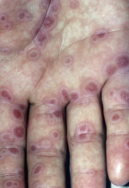
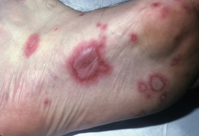
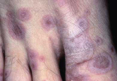
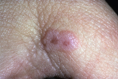
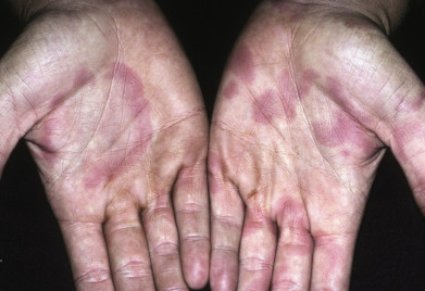
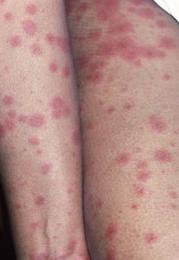
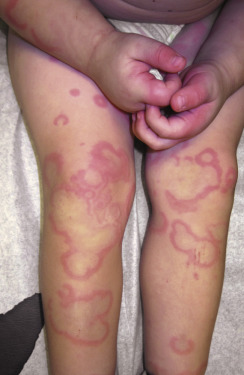
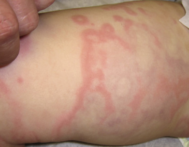
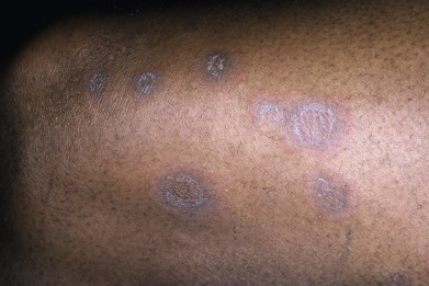
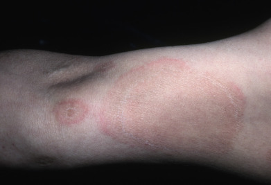
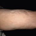
Many of the erythemas in this portion of the atlas share an annular and polycyclic morphology, including urticaria, erythema annulare centrifugum, and erythema gyratum repens, but only erythema multiforme demonstrates the true fixed target lesion. Noting whether skin findings are migratory or fixed is helpful in distinguishing common urticaria from many other more fixed lesions. Skin testing to demonstrate dermatographism is also helpful as a bedside technique to recapitulate urticaria in a susceptible patient. Ultimately, a skin biopsy may be needed to differentiate reactive neutrophilic dermatoses, such as Sweet syndrome (acute febrile neutrophilic dermatosis), from eosinophilic conditions, such as Wells syndrome (eosinophilic cellulitis). Tissue cultures are often needed when diagnosing pyoderma gangrenosum to rule out infectious etiologies such as bacteria, fungi, or atypical mycobacteria, depending on the clinical setting.
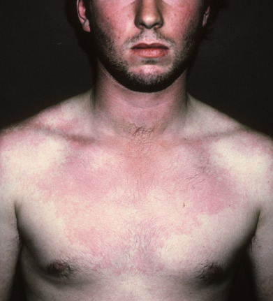
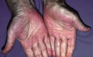
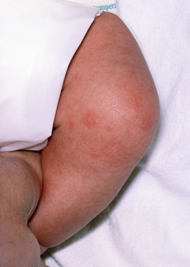
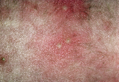
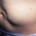
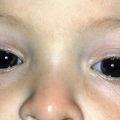
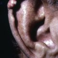
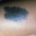
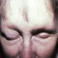
This portion of the atlas features examples of urticaria, urticarial lesions, erythemas, and angioedema.