Fig. 34.1
Epithelioid sarcoma. A slow-growing indurated subcutaneous tumor on the left arm of a 33-year-old man for 5 years (Courtesy of Cristina Cotruta, M.D. and Konstantinos Koutsioukis, M.D.)
Clinical Features
ES presents as a slowly growing dermal nodule that may rapidly ulcerate, a lobular subcutaneous tumor, or as a poorly defined deeply located mass. It may be asymptomatic or may cause paresthesia, pain, or muscular wasting. The classic type presents mainly in adolescents and young adults, while the proximal type affects mostly middle-aged or older adults. However, the neoplasm may occur at any age (range 11–93 years). Occurrence in children is uncommon. Males are more affected than females. The tumor measures less than 5 cm in diameter (range 1–30 cm). The time delay before diagnosis ranges from 2 to 96 months.
Classic-type ES typically occurs in the distal extremities, especially hand and forearm, but may also involve the leg, foot, and knee. It presents as a slowly growing, solitary or multiple, dermal or subcutaneous tumor, often accompanied by superficial ulceration, hemorrhage, and necrosis. The tumor has a tendency for extensive spread along blood vessels, nerves, and fascia, with appearance of satellite nodules in a sporotrichoid distribution. Proximal-type ES is less common and appears mainly in axial or proximal regions, including the limb girdles, pelvis, perineum, genitalia, mediastinum, and trunk. It presents as a deep, infiltrative mass that can extend along tendon sheaths or aponeuroses. ES may mimic other benign or malignant diseases, including perforating granuloma annulare, Dupuytren’s disease, and melanoma.
Pathology
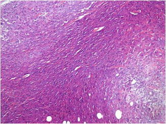
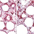
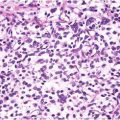
Classic-type ES is usually situated in the dermis and/or subcutis and has a nodular or diffuse outline. A “geographic” appearance with multiple nodules of intensely eosinophilic polygonal, epithelioid, or spindle-shaped cells associated with central necrosis is apparent even at scanning magnification (Fig. 34.2). Necrosis in the center of nodules may be a prominent feature and may lead to confusion with a granulomatous process (Fig. 34.3). At the periphery of the nodules, the tumor cells tend to be more spindle shaped (Fig. 34.4). The cells show variable pleomorphism and only a few mitotic figures (Fig. 34.5). Local infiltration along tendons, fascial planes, and neurovascular bundles is often present. These changes are more pronounced in advanced stages or in recurrent tumors and may not be evident in early lesions. The superficial tumors show less pleomorphism and a lower mitotic activity than deeply seated tumors. Proximal-type ES is characterized by sheets of large epithelioid cells, with vesicular nuclei and prominent nucleoli (Fig. 34.6). Cells with abundant glassy cytoplasm, eccentric nuclei, and prominent nucleoli (rhabdoid morphology) are frequently encountered. Some cases show a vague fibrinoid or myxoid pattern of degeneration. In a few cases, an angiomatoid or angiosarcoma-like pattern is seen. A “fibroma-like” variant has also been described. Focal calcification is an unusual feature.
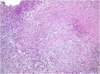
Fig. 34.2
The multinodular tumor with deeply eosinophilic appearance is located in the subcutaneous fat with minimal extension into the dermis
Fig. 34.3
There is extensive necrosis in the center of nodules which may lead to confusion with a granulomatous process