Enucleation and orbital implant placement


Table 73.1

Indications for surgery


Blind and painful eye
Sympathetic ophthalmia prophylaxis after uveal prolapse
Intraocular malignancy (e.g. choroidal melanoma, retinoblastoma)
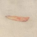
Phthisis bulbi with disfigurement and blind eye from multiple causes (post-traumatic, retinal detachment, end-stage glaucoma, endophthalmitis)


Table 73.2

Preoperative evaluation


Vision potential of eye
Pupillary response (reverse testing if globe and pupil are degenerated)
Motility
Eyelid and superior sulcus
Conjunctiva and fornices
Orbit (volume)
Intraocular pressure
Complete examination of fellow eye
Timing of injury
Oncologic work-up for cases of intraocular malignancy
Psychological status of patient (particularly for younger patients)


Introduction


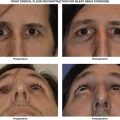
Enucleation is one of the oldest ophthalmic procedures and involves removal of the entire globe and portions of the optic nerve. The indications include blind and painful eye from a variety of causes, intraocular malignancy, and sympathetic ophthalmia prophylaxis after severe ocular trauma.


Importantly, the loss of an eye – especially a seeing eye – is strongly associated with depression and other psychological disturbances and, accordingly, referral to mental health professionals is often indicated.


During the evaluation, a careful examination is performed of the eye to be enucleated. The visual potential should be fully assessed, particularly in traumatic cases where some usable vision may remain. In cases where no view of the posterior pole is possible, a B-scan ultrasound should be performed to rule out occult intraocular tumor. The fellow eye must also be carefully evaluated and any pathology treated as the patient will be monocular. Patients are strongly advised to wear protective eyewear at all times to preserve the fellow eye.


The preference for evisceration ( Chapter 72 ) or enucleation varies between surgeons. With cases of known or suspected intraocular malignancy, enucleation is the standard of care, as complete pathologic examination is necessary. Obtaining a long optic nerve segment is particularly important in the management of retinoblastoma as this malignancy can spread via the optic nerve. Furthermore, a systemic oncologic evaluation should be performed in cases of intraocular malignancy.


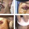
Restoring orbital volume after enucleation is paramount to achieving the best aesthetic and functional outcomes. A multitude of different orbital implants have been described over the past 50 years. The generally accepted consensus is to place the largest possible implant at the time of surgery. Currently used alloplastic implant materials at the time of writing include porous polyethylene (Medpor, Stryker Surgical, Kalamazoo, MI), hydroxyapatite (Bio-Eye, IOI, San Diego, CA) silicone, acrylic, and aluminum oxide bioceramic (FCI, Cedex, France).


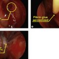
Fibrovascular integration of orbital implants lessens the chance of implant extrusion. Implants may also be wrapped with tissue such as fascia, autologous or donor sclera or bovine pericardium to decrease the risk of extrusion and to facilitate attachment of the extraocular muscles. The newer implants such as porous polyethylene do not require wrapping and the extraocular muscles can be directly attached to the implant.


Pegging of the implant can be performed to improve motility of the prosthetic but the high complication rates with this modification have largely limited its widespread use. Other complications related to enucleation include superior sulcus deformity, upper eyelid ptosis, and lower eyelid malpositions. Socket contracture is a challenging surgical condition and meticulous surgical technique at the time of enucleation can minimize this problematic condition.
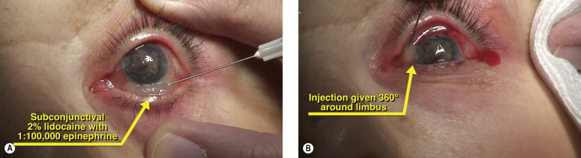
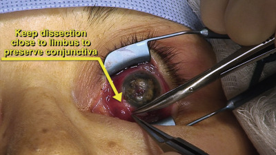
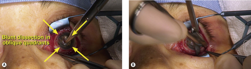
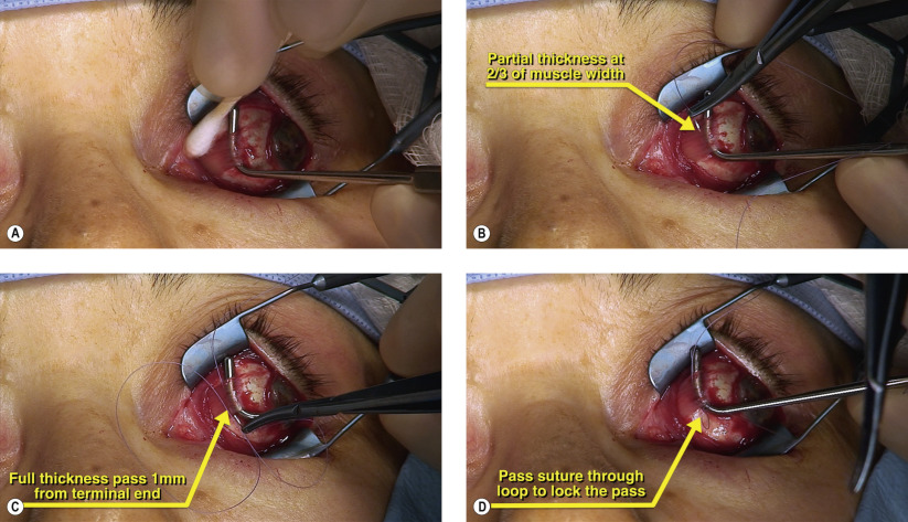


Surgical Technique


Figures 73.1A and 73.1B


Injection of local anesthetic

Enucleation is best performed under general anesthesia. Prior to the start of surgery, a “time out” is performed by all members of the team to confirm the correct eye for enucleation. Removal of the wrong eye is an unacceptable complication. If necessary, dilation can be performed to visualize intraocular pathology, particularly if there is no gross pathology evident on external examination of the globe. A corneal shield is placed on the fellow eye for protection. A retrobulbar injection provides maximal anesthesia and can facilitate hemostasis. To maximize hemostasis anteriorly, local anesthetic containing 1 : 100,000 epinephrine is given in a subconjunctival fashion around the limbus ( Figures 73.1A and 73.1B ). This local anesthesia also helps to hydrodissect the conjunctiva from Tenon’s capsule during the conjunctival peritomy.


Figure 73.2


Conjunctival peritomy

A wire eyelid speculum is placed to facilitate exposure. A 360° limbal peritomy is performed with Westcott scissors. During the peritomy, care should be taken to preserve the maximum amount of conjunctiva to ensure an adequate cul-de-sac postoperatively ( Figure 73.2 ). The blunt tips of the scissors are directed towards the sclera to prevent damage to the conjunctiva. In traumatic cases with gross disruption of normal anatomy, every effort should be made to preserve viable conjunctiva and avoid creating buttonholes in the tissues. Occasionally, scleral ruptures may be noted and these should be closed with 6-0 silk to minimize uveal extrusion and to maintain globe pressure during subsequent steps. Any dark, uveal tissues should be removed and irrigated away to lessen the theoretic risk of sympathetic ophthalmia.


Figures 73.3A and 73.3B


Exposure of extraocular muscles

Once the peritomy has been completed, Tenon’s capsule is dissected from the underlying sclera. Curved, blunt-tipped tenotomy scissors are placed with the closed tips hugging the sclera in the oblique quadrants ( Figure 73.3A ). The scissors are then spread widely as the instrument is withdrawn, bluntly widening Tenon’s capsule ( Figure 73.3B ). This is repeated multiple times in each oblique quadrant. Care is taken to not sever any of the extraocular muscle during the blunt dissection. Adequate dissection will facilitate exposure of the extraocular muscles in the subsequent step.


Figures 73.4A–D


Isolation and imbrication of medial rectus

After freeing conjunctiva and Tenon’s capsule from the underlying sclera, the rectus muscles are sequentially isolated and secured. Wide platform instruments such as the Von Graefe and Green muscle hooks are used to isolate the rectus muscles. In a non-traumatic eye, the medial rectus can be isolated first. When isolating the muscle, the hook is widely swept to ensure complete purchase of the entire muscle width. Once the muscle has been hooked, a cotton-tipped applicator is used to strip away the muscular capsule and expose the rectus fibers ( Figure 73.4A ). The muscles are secured with double-armed 6-0 Vicryl suture on a spatulated needle, passed in two-thirds width bite in a partial thickness fashion ( Figure 73.4B ). The terminal end of the suture is then passed full thickness and locked in a double whip stitch fashion ( Figures 73.4C and 73.4D ). Once the muscle is secured, scissors are used to disinsert the rectus muscle from sclera. When severing the rectus muscles, it is useful to leave a 2 mm stump of tendon so that a forceps, hemostat, or 5-0 silk suture can be placed for globe traction. The double-armed suture is then temporarily secured with a bulldog serrefine clamp.

Only gold members can continue reading. Log In or Register to continue

Stay updated, free articles. Join our Telegram channel

May 16, 2019 | Posted by in Reconstructive surgery | Comments Off on Enucleation and orbital implant placement

Full access? Get Clinical Tree

Get Clinical Tree app for offline access