Endoscopic dacryocystorhinostomy with osteotome


Introduction


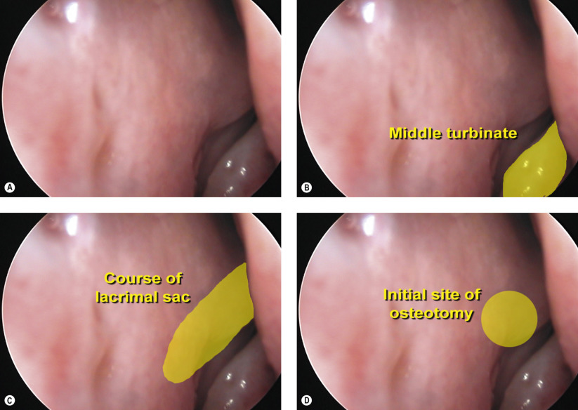
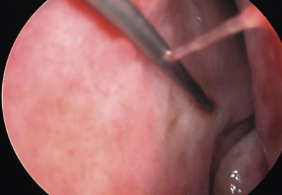
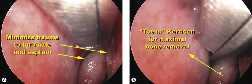
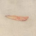
In this chapter, endoscopic dacryocystorhinostomy (DCR) with the use of an osteotome is presented. For indications and preoperative evaluation, refer to Chapter 53 . The osteotome is a useful adjunct during endoscopic DCR to rapidly remove the difficult-to-reach bone at the superior portion of the lacrimal sac fossa. Once the lacrimal sac has been exposed using the Kerrison ronguer, the 6 mm osteotome is directed vertically to fracture the frontal process of the maxilla along the lacrimal sac fossa. Once the vertical osteotomy has been completed, the osteotome is rotated horizontally to fracture the remaining thin lacrimal bone. The entire maxillary–lacrimal complex encompassing the lacrimal sac fossa can often be removed as a single unit.
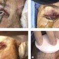
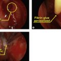


Surgical Technique


Figures 54.1A–D


Endonasal anatomy and surgical planning

In this endoscopic view, the middle turbinate can be clearly visualized ( Figures 54.1A and 54.1B ). Adequate nasal decongestion has been performed as evidenced by the appearance of the mucosa. Directly anterior to the middle turbinate is the course of the lacrimal sac, as shown in yellow ( Figure 54.1C ). The initial osteotomy site will be performed in this area ( Figure 54.1D ).


Figure 54.2


Injection of local anesthetic

Nasal decongestion is facilitated by packing neurosurgical cottonoids soaked in a 50/50 mixture of 4% lidocaine and oxymetazoline solution or 1 : 10,000 epinephrine (no cardiac contraindications) into the middle meatus. Further hemostasis is achieved by direct infiltration of lidocaine with epinephrine into the site of the initial osteotomy along the maxillary line using a 22-gauge, 3 inch spinal needle ( Figure 54.2 ).


Figures 54.3A and 54.3B


Creation of osteotomy

A 4 mm Kerrison ronguer is used to create the initial osteotomy. Care should be taken to avoid traumatizing the nasal septum or surrounding mucosa during all endonasal manipulations ( Figure 54.3A ). Nasal mucosa and bone overlying the lacrimal fossa are removed en-bloc. The footplate of the Kerrison ronguer should be squarely placed in a “toe-in” configuration to slightly infracture the lacrimal bone and to firmly engage the thicker maxillary bone within the lacrimal sac fossa ( Figure 54.3B ).

Only gold members can continue reading. Log In or Register to continue

Stay updated, free articles. Join our Telegram channel

May 16, 2019 | Posted by in Reconstructive surgery | Comments Off on Endoscopic dacryocystorhinostomy with osteotome

Full access? Get Clinical Tree

Get Clinical Tree app for offline access