Eczema
Overview
Despite being the most common inflammatory skin condition, eczema is the most confusing skin ailment for both patients and their nondermatologic health care providers. Eczema is very difficult to define. United States Supreme Court Justice Potter Stewart once said that he could not define pornography, but he knew it when he saw it. Such is the case with eczema, a condition that is best understood through repeated viewing.
The word eczema was coined by the Ancient Greeks to mean “a boiling out or over.” Conceivably, Greeks viewed certain rashes as boiling out or erupting from under the skin. As a case in point, the acute eczematous eruption of poison ivy often manifests with a fiery red color and a linear, blistered appearance, suggesting an acute boiling, bubbling, second-degree burn.
Terminologic confusion may also arise if the word dermatitis—a more generalized, often vague designation that refers to all cutaneous conditions with inflammation—is used synonymously with eczema or is coupled with it. In general, it is acceptable to use eczema and dermatitis interchangeably. Eczematous dermatitis, therefore, is somewhat redundant, although some might argue that the term is more inclusive than either word alone. The diversity of clinical images presented in this chapter is indicative of the protean clinical appearance of eczema.
Histopathology
On a microscopic level, an eczematous epidermis contains intercellular and intracellular fluid that appears in a spongelike formation (spongiosis). Vasodilatation of the dermis also occurs. These abnormalities result in the clinical manifestations of acute eczema: edema, erythema, vesicles, and bullae (e.g., from poison ivy).
Later, the epidermis thickens (acanthosis) and retains nuclei (parakeratosis), and an abundant cellular infiltrate develops in the dermis. These changes account for the scale and lichenification (see following) of chronic eczema (e.g., chronic lichenified atopic dermatitis).
Acute, Subacute, and Chronic Eczema
In reference to eczema, the designators acute, subacute, and chronic are somewhat arbitrary because they describe parts of a dynamic spectrum. A patient can present with lesions in any or all of the phases.
The following clinical presentations of eczema or associated conditions are discussed in this chapter:
 ATOPIC DERMATITIS (ATOPIC ECZEMA)
 CONTACT DERMATITIS AND DIAPER DERMATITIS
 CHRONIC HAND ECZEMA AND DYSHIDROTIC ECZEMA
 NUMMULAR ECZEMA
 LICHEN SIMPLEX CHRONICUS
 PRURIGO NODULARIS
 ASTEATOTIC ECZEMA
 NEUROTIC EXCORIATIONS
 NONSPECIFIC ECZEMATOUS DERMATITIS
 SEBORRHEIC DERMATITIS
 STASIS DERMATITIS
 2.1 Acute allergic eczematous eruption of poison ivy. The red color and linear blistered appearance suggest an acute “boiling,” bubbling, second-degree burn. |
 2.2 Subacute eczema. The crusts, scales, and erythema of subacute eczema are less intense than those seen in acute eczema. |
Acute eczema is manifested by itchy erythematous patches, plaques, or papules that may become “juicy” and develop into vesicobullous lesions (Fig. 2.1). Alternatively, acute eczema may originate and continue as a less florid, nonvesicular, erythematous eruption.
Subacute eczema is an intermediate stage between acute and chronic eczema. The term has little clinical value. It is best simply to be aware that acute oozing lesions dry into crusts (scabs; Fig. 2.2), and they can later develop scales that overlie an erythematous base. Subacute eczema may become chronic, resolve spontaneously, or resolve with treatment.
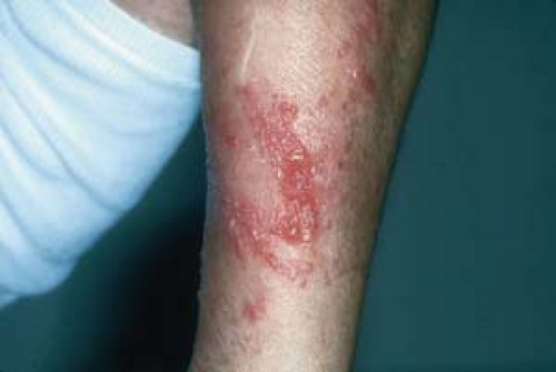
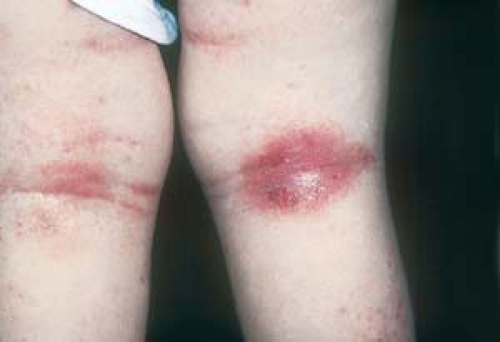
Chronic eczema is also known as chronic eczematous dermatitis. Its hallmark is lichenification—plaque with an exaggeration or hypertrophy of the normal skin markings. Lichenification resembles the bark on a tree trunk; or, as implied, the skin appears lichenlike. (Fig. 2.3). In addition, scale and hemorrhagic crusts can result from scratched or drying vesicles. Older lesions may exhibit postinflammatory pigment alterations (PIPA), i.e., hyperpigmentation and/or hypopigmentation (Fig. 2.4).
 2.3 Chronic eczematous dermatitis. This patient shows lichenification, an exaggeration of skin markings, which was caused by repeated scratching. |
 2.4 Chronic eczematous dermatitis. This lesion shows no evidence of active inflammation. Lichenification and postinflammatory hyperpigmentation are apparent. |
Atopic Dermatitis (Atopic Eczema)
See also Chapter 27, “Special Considerations in the Skin of Pediatric and Elderly Patients.”
Basics
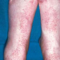
Atopic dermatitis, also known as atopic eczema or endogenous eczema, is the most commonly seen type of eczema (Figs. 2.5, 2.6, 2.7, 2.8 and 2.9). It is a chronic, inflammatory, itchy skin condition with an unpredictable course of flares and remissions that affects an estimated 5% to 10% of the United States population. Atopic dermatitis is the most frequently seen skin condition among patients of Asian descent.
By definition, atopic dermatitis occurs in association with a personal or family history of hay fever, asthma, allergic rhinitis, sinusitis, or atopic dermatitis itself. A probing history taking is often necessary to uncover symptoms of atopy. For example, patients should be asked whether they or their family members are allergic to pollen, dust, house dust mites, ragweed, dogs, or cats. Inquiries should be made about chronic recurrent symptoms that suggest atopy, such as nasal pruritus and rhinitis, rhinorrhea, paroxysmal sneezing, or itchy or irritated eyes. A personal or family history of allergies to multiple medications is also important. Furthermore, secondary relatives (aunts, uncles, cousins, and grandparents) may have an atopic predisposition.
Most cases begin in childhood (often in infancy); however, atopic dermatitis may start at any age. The disease frequently remits spontaneously—reportedly in 40% to 50% of children—but it may return in adolescence or adulthood and possibly persist for a lifetime. Traditionally, patients and their families were advised that children “will grow out of eczema”; however, this optimistic prognosis is not always realized.

2.5 Atopic dermatitis. The cheeks are a typical location in an infant.

2.6 Atopic dermatitis. Lesions are widespread in this infant.

2.7 Atopic dermatitis. Antecubital involvement in a 2-year-old child.
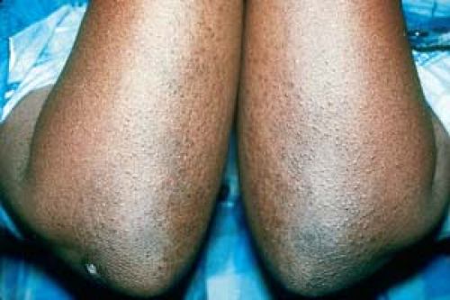
2.8 Atopic dermatitis. Popliteal involvement is apparent in this toddler.
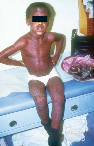
2.9 Atopic dermatitis, generalized. Note the marked postinflammatory hyperpigmentation in this African-American child.
Children with asthma—an increasing population in the inner cities of the United States—appear to have a higher prevalence of atopic dermatitis than do other children; atopic dermatitis often manifests in asthmatic children in a more extensive and chronic form. African-Americans and Asians, particularly those living in an urban setting, tend to develop atopic dermatitis at an earlier age. Severe eczema in childhood portends a worse prognosis in adulthood.
Atopic dermatitis can present with a wide spectrum of severity. Some patients may have only a mild, recurrent, localized, itchy rash on “dry” or “sensitive” skin; others may experience a more severe, extensive eruption that can be accompanied by unremitting pruritus, sleepless nights, secondary cutaneous bacterial or viral infections, embarrassing alligatorlike lichenification, and, rarely, an exfoliative erythroderma. Many patients with atopic dermatitis have multiple accompanying atopic ailments, as mentioned earlier.
In addition to the physical discomfort of atopic dermatitis, patients may suffer from embarrassment about the appearance of lesions. Psychosocial problems, such as poor self-image, anger, and frustration, may lead to depression and social isolation.
Common Myths About Eczema
Myth: Soy formulas improve eczema in infants.
Fact: Food allergies that induce eczema are rare. Urticaria (hives) is the usual skin manifestation of a food allergy.
Myth: Infants and children who have atopic dermatitis should be bathed infrequently.
Fact: The advantages of bathing far outweigh its disadvantages (see following).
Myth: Laundry detergents are a common cause of eczema.
Fact: Very rarely is this the case. Most detergents are rinsed out, with very little soap remaining to cause a rash.
Myth: Potent topical steroids should not be used on infants or young children.
Fact: Topical steroids, when used properly, are quite safe. Not using them can lead to undertreatment that can last for an indeterminate period of time.
Pathogenesis
Atopic dermatitis is an inherited type I (immunoglobulin E–mediated) hypersensitivity disorder of the skin. In comparison with normal skin, atopic skin tends to be more prone to irritation, dryness, barrier abnormalities, and infection; it is also more likely to be negatively influenced by emotional stress.
The intense itching of atopic dermatitis is presumed to be produced by the release of vasoactive substances from sensitized mast cells and basophils in the dermis. The itching may be initiated by external agents such as woolen or synthetic fabrics; certain foods; alcohol; and overexposure to dry, cold weather or to very hot, humid conditions that predispose to sweating. Less commonly, pruritus has been reported to be triggered by house dust mites. There is considerable debate about whether atopic dermatitis is primarily an allergen-induced disease or, rather, an inflammatory skin disorder found in association with respiratory allergy or other atopic symptoms. Individuals find that an allergic condition is permanent, whereas they often outgrow atopic dermatitis; this supports the latter explanation.
Even though atopic dermatitis frequently remits spontaneously, patients, their families, and their health care providers—in particular, pediatricians and allergists—often relentlessly search for external sources that patients can avoid or eliminate from their environments. Avoidance of milk products and food preservatives and extreme dietary restrictions are not only very difficult to maintain on an ongoing basis, but they may also incite developmental problems in growing children; furthermore, they rarely, if ever, offer a cure.
Description of Lesions
Although the character and distribution of the skin eruption tend to vary according to the patient’s age, the different phases of atopic dermatitis are not always clearly distinct.
Any or all manifestations of atopic dermatitis may exist in a single patient.
Infantile Phase
In patients aged 2 months to 2 years, the face (particularly the cheeks; see Fig. 2.5) scalp, chest, neck, and extensor extremities are most often involved. The eruption may become generalized (see Fig. 2.6). In many cases, atopic dermatitis first manifests with severe “cradle cap” or severe recalcitrant intertriginous (groin, neck, axillae) rashes.
Childhood Phase
Lesions seen in children aged 2 through 12 years tend to become lichenified because of repeated rubbing and scratching.
Lichenification occurs more commonly in Asian and African-American patients than in Caucasian patients (see Fig. 2.9).
The hallmark of atopic dermatitis is pruritus; children who have atopic dermatitis are typically very “busy” and cannot often sit quietly because of the pruritus.
Distribution of Lesions
Lesions tend to occur symmetrically, with a characteristic distribution in the flexural folds: the antecubital and popliteal fossae and the neck, wrists, and ankles.
Lesions may also occur on the eyelids, lips, scalp, and behind the ears.
Adolescent and Adult Phase
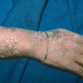
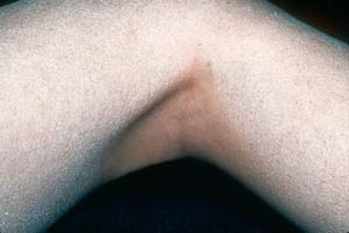
In adolescents and adults, lichenified plaques are generally prominent and tend to blend into surrounding normal skin. Postinflammatory hyperpigmentary and hypopigmentary changes may be seen (Fig. 2.10). Alternatively, lesions may consist of small, itchy, erythematous papules (e.g., follicular eczema; Fig. 2.11) or vesicles on the hands (e.g., dyshidrotic eczema; see below).
 2.10 Atopic dermatitis. In this adult, the lichenified plaques tend to blend into the surrounding normal skin. Postinflammatory hyperpigmentation is seen here. |
 2.11 Atopic dermatitis, follicular eczema. Atopic dermatitis of the hair follicles. Note the gridlike pattern of follicular papules. This is a common presentation in African-American patients. |
 2.12 Atopic dermatitis. Lesions in this patient resemble both nummular and asteatotic eczema (see Figs. 2.40 and 2.52). |
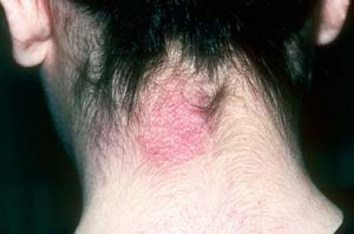 2.13 Atopic dermatitis, lichen simplex chronicus. The nape of the neck is a common site of involvement. |
Distribution of Lesions
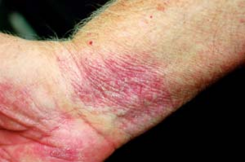
The distribution of lesions may be similar to that seen in childhood (i.e., in the flexural folds). However, lesions may also appear in extensor locations: the dorsa of the hands, wrists, shins (Fig. 2.12), ankles, and feet, and the nape of the neck (Fig. 2.13). On the other hand, lesions may be limited to the lips (atopic cheilitis; Fig. 2.14); eyelids; vulvar or scrotal areas (Figs. 2.15 and 2.16, respectively); or hands (as in chronic hand eczema or dyshidrotic eczema; see below), which may be the only features of atopic dermatitis in some adults. Nail dystrophy occurs when the proximal nail fold and the underlying nail matrix (root) are involved (Fig. 2.17).
Other Clinical Aspects
Additional associated features and findings that are clues to the diagnosis of atopic dermatitis include the following:
Persistent xerosis, or dry, “sensitive” skin.
Dennie–Morgan lines. These comprise a characteristic double fold that extends from the inner to the outer canthus of the lower eyelid (Fig. 2.18).
“Allergic shiners.” This term refers to a darkened, violaceous, or tan coloring in the periorbital areas. Along with Dennie–Morgan lines, this dark coloring may be an instant clue to the diagnosis of atopic dermatitis (Fig. 2.19).
Hyperlinear palmar creases (Fig. 2.20).
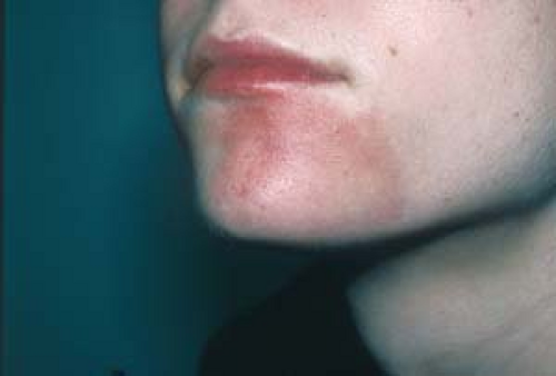
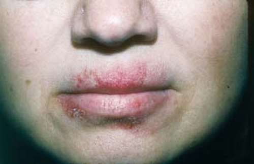 2.14 Atopic cheilitis (atopic dermatitis of the lips). Note the lichenification and the ill-defined outline of the vermilion border of the upper lip. |
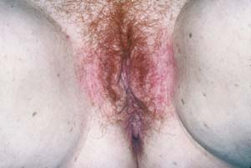 2.15 Atopic dermatitis limited to the vulvar and inguinal areas. This eruption was initially diagnosed and treated as a fungal infection by the patient’s health care provider. |
 2.16 Atopic dermatitis (lichen simplex chronicus) limited to the scrotum. This patient was also initially thought to have tinea cruris. Note the lichenification. |
 2.17 Atopic dermatitis. This patient’s middle fingernails show dystrophic changes, transverse ridging, and cuticle loss solely on those fingers where eczema is present in the proximal nail folds. |
 2.18 Atopic dermatitis, periorbital. Note lichenification and the characteristic double fold (Dennie-Morgan line) that extends from the inner to the outer canthus of the lower eyelid and the “allergic shiners,” the darkening color of the periorbital areas. |
 2.19 Atopic dermatitis, periorbital. The presence of “allergic shiners”—a darkened or violaceous hue, as in this child—is a clue to the diagnosis of atopic dermatitis. |
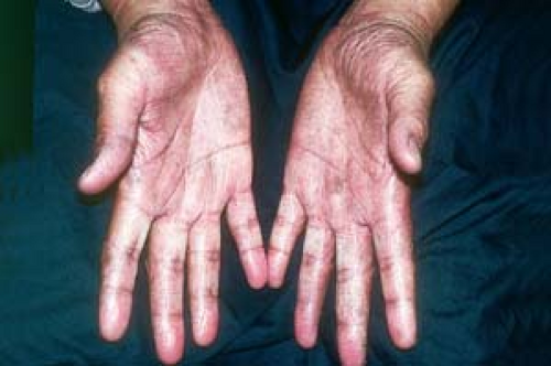 2.20 Atopic dermatitis. Hyperlinearity of the palms is evident here. |
 2.21 Ichthyosis vulgaris. Lesions resemble fish scales. Note the characteristic sparing of the popliteal creases. |
 2.22 Keratosis pilaris. This teenager has tiny, rough-textured, red, follicular papules on his lateral upper arms. |
Other associated dermatoses include the following:
Ichthyosis vulgaris. This condition is frequently associated with atopy. Lesions, which are most apparent on the shins, resemble fine fish scales. Characteristically, the flexor creases are spared in this condition (Fig. 2.21).
Keratosis pilaris. These tiny, horny, rough-textured, whitish or red, follicular papules or pustules occur most often during adolescence. Most commonly, keratosis pilaris is noted on the deltoid and posterolateral upper arms, the upper back and thighs, and the malar area of the face. It is frequently confused with acne (Fig. 2.22).
Clinical Manifestations and Possible Complications
Pruritus may interfere with sleep. Itching is increased by repeated scratching and rubbing, which leads to lichenification, oozing, and secondary bacterial infection (impetiginization, or “honey-crusted skin”).
Secondary infection with Staphylococcus aureus may trigger relapses of atopic dermatitis.
Secondary infections with herpes simplex virus may result in eczema herpeticum (Kaposi’s varicelliform eruption), which is more commonly seen in childhood.
Diagnosis
Diagnosis of atopic dermatitis depends on excluding conditions such as fungal infections, seborrheic dermatitis, psoriasis, scabies, contact dermatitis, ichthyosis, and cutaneous T-cell lymphoma.
The diagnosis of atopic dermatitis is generally not difficult, especially in patients with an atopic history in whom the following causes of eczema or eczemalike eruptions have been excluded.
Contact Dermatitis
(See later in this chapter).
Determine whether the patient was exposed to a substance that could cause contact dermatitis. The location of the lesions may suggest an external cause.
Scabies
(See Chapter 20, “Bites, Stings, and Infestations.”)
A history of exposure is important in diagnosing scabies.
Symptoms are present in other household members.
Characteristic distribution (e.g., in the webs between the fingers and on the flexor wrists) can mimic that of eczema.
A positive scabies scraping is diagnostic of scabies.
Psoriasis
See Chapter 3, “Psoriasis.”
Lesions generally appear on extensor locations—the elbows, knees, and other large joints—rather than on flexor creases.
Patients may have a positive family history of psoriasis.
Usually, psoriasis is less pruritic than eczema.
Psoriatic lesions tend to be clearly demarcated from normal surrounding skin, and the scale of psoriasis tends to be thicker and micaceous in appearance. However, psoriasis may at times be clinically indistinguishable from atopic dermatitis.
Tinea Pedis, Corporis, Manuum, and Capitis
(See Chapter 7, “Superficial Fungal Infections.”)
A positive potassium hydroxide (KOH) test or fungal culture result indicates these conditions.
Topical Steroid Therapy
See also “Introduction: Topical Therapy.”
General Principles
The application of an appropriately chosen topical steroid usually results in prompt improvement in most patients with atopic dermatitis.
Topical steroids should be used only as short-term therapy, if possible, and only to treat active disease (i.e., with itching and erythema).
Topical steroids should not be used for prevention of future lesions or for cosmetic concerns, such as postinflammatory hyperpigmentation.
“Stronger” is often preferable to “longer” in the use of topical steroids, because long-term application is more often associated with side effects.
Without question, the use of a superpotent topical steroid is preferable to the administration of a systemic steroid, with its potential side effects.
When the condition is under control, the frequency of application and the potency of the topical steroids are reduced (“downward titration”), or the agents are discontinued.
Face and Body Folds
For the face and intertriginous regions (the axillae and inguinal creases are areas that are “naturally” occluded), treatment should be initiated with a low-potency (class 7) cream or ointment, such as over-the-counter (OTC) hydrocortisone 0.5% or 1%.
In more severe cases of atopic dermatitis, initial therapy may be with a more potent steroid, followed by a less potent preparation for maintenance. For example, a higher-potency (class 5) agent, such as hydrocortisone valerate 0.2% cream, could be used for 2 or 3 days, followed by a lower-potency (class 6) agent, such as desonide 0.05% or OTC hydrocortisone 0.5% to 1%, which is then used for maintenance as needed.
Body
For nonintertriginous areas of the body, treatment can be initiated with a mid-strength (class 4) cream or ointment, such as triamcinolone acetonide 0.1%, or a mid-strength (class 3) agent, such as fluocinonide 0.05%. Even a superpotent (class 1) agent, such as clobetasol 0.05%, may be used for limited periods (no more than 2 weeks) until control is achieved. Then therapy may be switched to a lower-potency (class 5) agent, such as hydrocortisone valerate 0.2%. Fluticasone 0.05% cream (Cutivate) is another lower-potency agent that has been shown to be effective as maintenance therapy when it is used twice weekly.
Ointments are helpful for dry skin. Because of their occlusive properties, they are more lubricating than other formulations. They also tend to be less irritating and less sensitizing. Their popularity increases in colder, drier weather. For patients with widespread skin involvement, a 1-lb jar of triamcinolone acetonide cream or ointment 0.1% is quite economical.
Infection
If necessary, topical steroids can be given concurrently with systemic antibiotics. This combination is sometimes helpful if evidence of coexisting staphylococcal or streptococcal infection (“impetiginization”) of the skin is present. Oral antibiotics such as cephalexin or dicloxacillin are commonly used. Also, tetracycline derivatives, which may be prescribed in patients over 10 years of age, are effective.
During flare-ups, the acute, open, weeping, crusted lesions that develop from infection or scratching may be treated with a drying antibacterial agent, such as Burow’s solution, before topical steroids are applied.
Clorox bleach—1 to 2 teaspoonfuls per tubful of water, which can be increased up to 6 teaspoonfuls added to daily bath water—is a quite effective and inexpensive alternative to topical and oral antibiotics.
Another topical option is topical 2% mupirocin cream (Bactroban) or ointment (Centany).
Topical Immunomodulator Therapy
Currently, these drugs are indicated for the short-term or intermittent treatment of atopic dermatitis and are considered as second-line therapy when conventional therapies are inadvisable, ineffective, or not tolerated. Photoprotection is advised when using these agents, although no evidence so far has implicated them in promoting skin cancer.
For information regarding the long-term safety of these agents, see “Introduction: Topical Therapy.”
Protopic ointment (tacrolimus) is a nonsteroidal immunomodulator that has been shown to reduce the symptoms of atopic dermatitis. It is used as an alternative to topical steroids, particularly when the eruption involves the face or intertriginous areas, such as the axillae and groin, where the long-term use of high-potency steroids is limited. Protopic has potential as a topical steroid–sparing agent.
When applied twice daily, Protopic may cause transient side effects, such as burning and itching. This burning and itching may result in patients’ refusal to continue applying the medication.
It is available as an ointment in 0.1% and 0.03% concentrations.
The 0.1% concentration has been approved for the treatment of atopic dermatitis in adults.
The 0.03% concentration is designated for short-term and intermittent long-term treatment of atopic dermatitis in children (age 2 years and older) and in adults.
Elidel cream (pimecrolimus), in a 1% cream base, is less likely to be irritating, and it is not as greasy as Protopic ointment.
Other Therapeutic Measures
Oral H1 antihistamines such as diphenhydramine (Benadryl) and hydroxyzine (Atarax) probably do not reduce itching, but they are sometimes useful as inducers of sleep, an unacceptable side effect for many patients.
Oozing, exudative lesions may be soothed and dried with Burow’s solution or by bathing in a tub with antipruritic emollients, such as an Aveeno oatmeal bath preparation.
Sun exposure, ideally in the early morning and late afternoon—when humidity is lowest—may significantly improve atopic dermatitis in selected patients.
When a patient does not have access to natural sunlight, phototherapy with ultraviolet B rays and, less commonly, ultraviolet A rays, is often very effective for widespread skin involvement.
Less commonly used today, tar baths, as well as tar preparations formulated as ointments, pastes, and gels, may be used concurrently with topical steroids or alternated with them. (Before the advent of topical steroids, tar preparations were the mainstay of treatment.)
Emotional stress in patients or in their families may contribute to atopic dermatitis. Measures to reduce stress include support groups and family psychotherapy.
Before resorting to systemic steroids, a “soak and smear” regimen may be used. (See Introduction, “Topical Therapy.”)
Systemic steroids, immunosuppressive therapy with systemic agents such as cyclosporine, or short-term hospitalization is sometimes necessary in patients with severe unresponsive generalized atopic dermatitis.
For patients with secondary herpes simplex infection (Kaposi’s varicelliform eruption), oral antiviral therapy and possibly hospitalization may be required.
Unsuccessful Treatments
Treatments that do not seem to improve atopic dermatitis include vitamins, mineral or dietary supplements, and other nutritional supplements.
Topical ointments that include diphenhydramine and doxepin are potential contact allergens and thus are contraindicated.
General Management
Bathing
How often the person who has atopic dermatitis should bathe has been the subject of controversy and misunderstanding. There are many reasons not to restrict frequent bathing:
Bathing removes crusts, irritants, potential allergens, and infectious agents.
Bathing provides pleasure and reduces stress.
Bathing hydrates the skin and allows better delivery of corticosteroids and moisturizers.
The addition of Clorox bleach to bath water (“bleach baths”) can be effective (see earlier discussion) for oozing or infected atopic dermatitis.
Bathing Tips
Mild, moisturizing soaps such as Dove or nonsoap cleansers such as Cetaphil Lotion should be used.
The patient should be cautioned not to use soap on lesional skin (many people are erroneously led to believe that scrubbing with “good soaps” may actually help inflamed skin).
Excessive bathing that is not followed immediately by application of a moisturizer tends to dry the skin.
Excessive toweling and scrubbing should be avoided.
Prevention of Atopic Dermatitis
The following measures may help the patient to avoid or reduce exposure to triggers such as dry skin, irritants, overheating and sweating, and allergens.
Dry skin: Moisturizers, particularly in the dry winter months, should be applied immediately after bathing to “trap” water in the skin. However, in warm climates or in the summer, moisturizers may actually be irritating or may interfere with healing.
Suggested ointments: Vaseline Petroleum Jelly, Aquaphor
Suggested creams and lotions: Eucerin, Cetaphil, Lubriderm, Curel, Moisturel
Barrier repair: Atopiclair, MimyX, and Epiceram are multiple-ingredient prescription, nonsteroidal barrier creams that are applied two or three times per day. Their efficacy as topical steroid–sparing agents has been suggested by several studies. The barrier cream and lotion CeraVe can be purchased OTC.
Irritants: Nonirritating fabrics, such as cotton, should be worn. Wool clothing may induce itching.
Overheating and sweating: Excess dryness or humidity should be avoided. An air conditioner or humidifier in a child’s bedroom may help to avoid the dramatic changes in climate that may trigger outbreaks. (Unfortunately, humidifiers do not seem to help very much.)
Allergens: Some evidence indicates that the environmental elimination of certain airborne substances, such as house dust mites, may bring some lasting relief. A great deal of controversy surrounds the influence of dietary manipulation and the value of skin testing and hyposensitization on the course of atopic dermatitis. Although some foods may provoke attacks, elimination of them rarely brings a lasting improvement or cure. Skin tests and allergy shots may actually provoke attacks of atopic dermatitis.
Topical steroids should be applied only to active disease (inflamed skin).
When topical steroids are applied immediately after bathing, their penetration and potency are increased.
Low-potency topical steroids are recommended for use on the face and in skin folds, such as the perineal area and underarms.
A very small number of children show some clinical improvement after measures to control house dust mites are instituted.
“Soak and smear” therapy can often be substituted for, or limit the use of, systemic steroids.
There are primarily two causes of eczema: one comes from the outside (contact dermatitis), and the other comes from the inside (atopic dermatitis).
Patients and their parents, caregivers, and teachers should be educated about the manifestations and management of atopic dermatitis.
The “gooiest” and cheapest moisturizer is petrolatum.
The National Eczema Association can be contacted at:
415-499-3474 or 800-818-7546
4460 Redwood Highway, Suite 16-D, San Rafael, CA 94903-1953
info@nationaleczema.org or http://www.nationaleczema.org
Contact dermatitis
 2.23 Irritant contact dermatitis. The localized erythema on this boy’s face was caused by irritation from benzoyl peroxide in an acne preparation. |
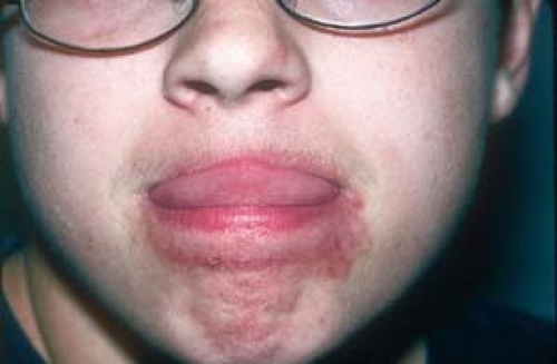 2.24 Irritant contact dermatitis. The erythema and scaling are obviously secondary to this child’s habit of licking her lips. |
 Irritant contact dermatitis. Chronic irritation from a stoma was the cause of this reaction. |
Contact dermatitis is an inflammatory reaction of the skin that is caused by an external agent. The appearance of the eruption and a careful history often give clues to the offending agent. The two types of contact dermatitis are irritant and allergic.
Irritant Contact Dermatitis
Also known as nonallergic contact dermatitis, irritant contact dermatitis (ICD) is an erythematous, scaly, sometimes eczematous eruption that is not caused by allergens (Figs. 2.23 and 2.24). A direct toxic reaction to rubbing, friction, or maceration, or to exposure to a chemical or thermal agent, the severity of ICD depends on the concentration of the irritant, its thermal energy, its abrasiveness, and the duration of exposure, among other factors. ICD may occur in anyone.
The eruption of ICD is confined to the areas of exposure, as exemplified by diaper rash, “dishpan hands,” and reactions that occur under an adhesive dressing (Fig. 2.25) or where a topical medication was applied. Examples of irritants include alkalis, acids, solvents, soaps, detergents, and numerous chemicals found in the home and workplace that damage the skin after repeated contact. Patients who have atopic dermatitis are more likely to develop ICD as a result of their inherent skin sensitivity and defective barrier function.
Diagnosis, when not clearly evident, is based on a careful history and the ruling out of allergic contact dermatitis (ACD), which is discussed later. Management is fairly simple: Patients should be told to avoid the offending agent or to minimize contact with it.
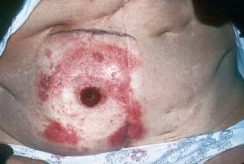
Diaper Dermatitis (Diaper Rash)
Basics
A common example of ICD is diaper dermatitis (diaper rash), referred to as “napkin dermatitis” in the United Kingdom. Diaper dermatitis applies to eruptions that occur in the area covered by a diaper. It can first present as early as the first few weeks of life or occur any time when diapers are worn.
Irritant diaper dermatitis is by far the most common rash in infancy, but it is not restricted to that age group because it can affect persons of any age group who wear diapers, such as incontinent patients.
Pathogenesis
Diaper dermatitis is essentially the result of overhydration of the skin that is irritated by chafing, by soaps and detergents, and by prolonged contact with urine and feces. The ammonia that is produced as a breakdown product of urea by bacteria in feces is considered an exacerbating factor.Related posts:

Stay updated, free articles. Join our Telegram channel

Full access? Get Clinical Tree