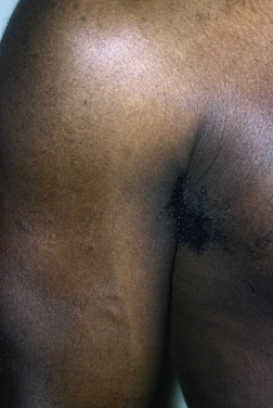
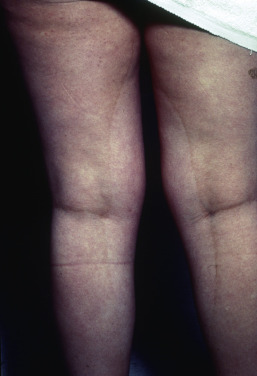
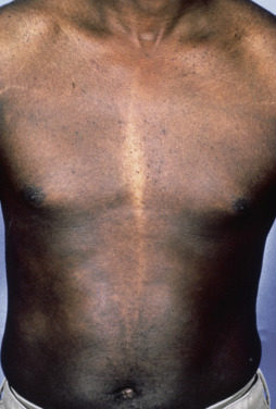
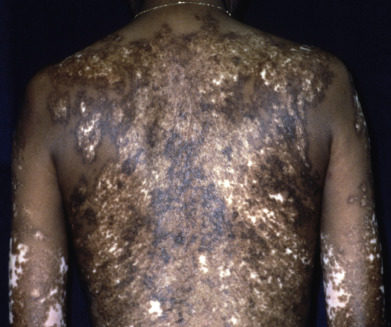
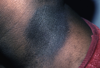
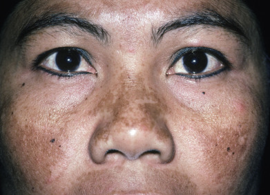
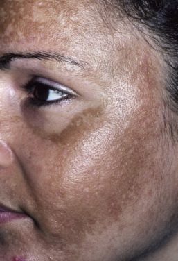
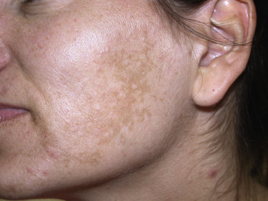
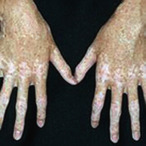
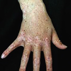
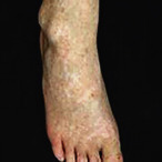
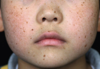
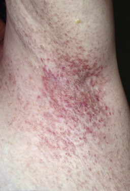
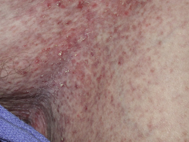
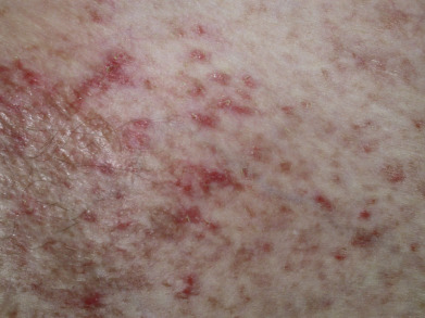
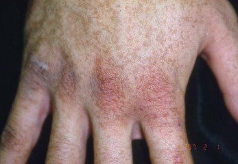
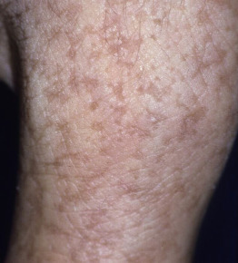
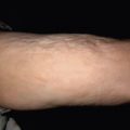
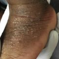
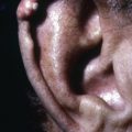
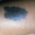
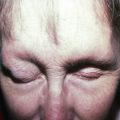
Pigmentary disorders of the skin may present with depigmentation, hypopigmentation, or hyperpigmentation. The color, morphology, and distribution of the lesions help to suggest the correct diagnosis. Vitiligo may be segmental (blaschkoid), acral (lips and tips), or truncal in distribution. Each presentation has implications for treatment. The lesions of vitiligo must be distinguished from those of pityriasis alba, hypopigmented mycosis fungoides, leprosy, and the confetti-like depigmentation and ash-leaf macules of tuberous sclerosis. Hyperpigmented lesions cover a similar spectrum of disorders from lichenoid dermatoses to melasma and the café-au-lait macules of neurofibromatosis. This section of the atlas will focus on disorders of pigmentation not found in other chapters of the book, including vitiligo, piebaldism, pigmentary demarcation lines, melasma, and Galli-Galli disease. A Wood’s lamp is of value in these disorders, as accentuation of the pigment suggests epidermal pigment alternation, whereas a decrease in the appearance of the pigmentary anomaly suggests dermal pigment deposition. Topical treatments can address many forms of epidermal dyspigmentation, whereas laser treatment is often more appropriate for the treatment of dermal pigment. When the clinical examination fails to provide a specific diagnosis, addition of a directed history or biopsy may be required. Fontana staining indicates the presence of melanin, and immunostains such as Mart-1 and Sox-10 confirm the pattern and distribution of melanocytes within the lesion.
Related posts:

Stay updated, free articles. Join our Telegram channel

Full access? Get Clinical Tree


