Disorders of the Mouth, Lips, and Tongue
 Common manifestations include:
Erosions, blisters, fissures, and ulcers
Erosions and ulcers of the tongue
Whitish plaques
Pigmentary changes
Neoplasms
Cystic lesions
Normal variants
Overview
Oral mucous membrane lesions are often clues to the presence of systemic illnesses, such as acquired immunodeficiency syndrome (AIDS), syphilis, and systemic lupus erythematosus. They may also be helpful in diagnosing dermatologic conditions, including lichen planus and pemphigus. Lesions such as “canker sores” are often seen as isolated phenomena but may also be an accompaniment or a precursor to a symptom complex, such as that seen in Behçet’s syndrome or ulcerative colitis.
It is often difficult to make a clinical diagnosis in the oral cavity. Lesions may resemble normal variants, or they may look like each other.
Inflammatory oral lesions—particularly those that are vesicobullous—rarely remain intact and unruptured. Instead, such lesions usually become erosions and ulcers by the time clinicians see them, adding to the difficulty of diagnosis.
Erosions, Blisters, Fissures, and Ulcers
Aphthous Stomatitis
Basics
Commonly known as “canker sores,” aphthous stomatitis lesions (aphthous ulcers) are a common, recurrent problem consisting of shallow erosions of the mucous membranes.
They are seen in children and adults and appear to be more common in women than men.
Aphthous stomatitis has no known cause, but an immune mechanism is considered the most likely contributory factor.
Patients often ascribe recurrences to psychologic stress or local trauma.
Women may correlate them with their menstrual cycle.
Most cases heal spontaneously, only to recur unexpectedly.
Oral erosions that are indistinguishable from aphthous stomatitis are sometimes seen in primary herpes simplex virus (HSV) infections.
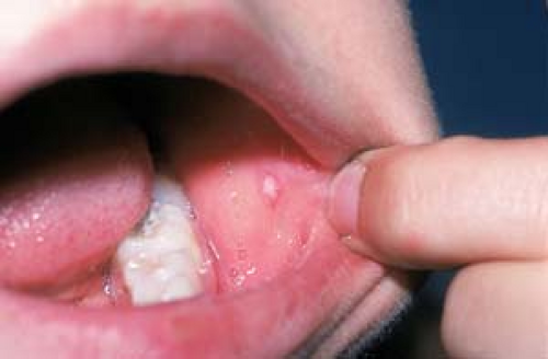 12.1 Aphthous stomatitis. A small punched-out erosion has a bright red margin surrounding a gray-white or yellowish center. |
Description of Lesions
The lesions of aphthous stomatitis are small (2 to 5 mm), shallow, well-demarcated, punched-out erosions.
Lesions typically have a ring of erythema, with a gray or yellowish center (Fig. 12.1).
Distribution of Lesions
Lesions may be seen on the buccal, labial, and gingival mucosa, as well as on the tongue (see Fig. 6.27).
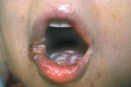 12.2 Aphthous stomatitis in Behçet’s syndrome. Large, painful erosions and extensive aphthae are evident on the lips and tongue of this woman. |
Clinical Manifestations
The lesions are usually painful.
They tend to heal in 4 to 14 days, a duration similar to that of HSV lesions.
Patients with human immunodeficiency virus (HIV) or Behçet’s disease may develop aphthous stomatitis lesions that are larger and more persistent, painful, and extensive than those seen in other patients (Fig. 12.2).
Diagnosis
The diagnosis is made clinically.
Most intraoral ulcers in immunocompetent patients are canker sores, not HSV lesions.
The presence of persistent or large painful aphthae broadens the differential diagnosis to include:
Primary HSV infection
Autoimmune bullous diseases such as pemphigus vulgaris and bullous pemphigoid
Recurrent erythema multiforme
Systemic lupus erythematosus
Cyclic neutropenia
Erosive oral lichen planus
Ulcerative colitis
Behçet’s disease
Therapeutic options for aphthous stomatitis lesions include:
Symptomatic therapy with topical anesthetic viscous lidocaine (Xylocaine) may help.
Vanceril (beclomethasone dipropionate) aerosol can be sprayed directly on lesions.
Superpotent topical steroids are applied directly to lesions and held there by pressure with a finger.
Tetracycline suspension (250 mg/tsp); patients should “swish and swallow.”
Diphenhydramine (Benadryl) suspension; patients are to “gargle and spit.”
Tacrolimus ointment (Protopic) 0.1% and (Elidel) pimecrolimus cream 1% applied at bedtime may accelerate healing.
Silver nitrate, applied directly to lesions, also can promote healing.
Intralesional corticosteroid injections or a brief course of systemic corticosteroids are effective in reducing pain and healing lesions in patients with large, persistent, painful ulcers.
Recently, thalidomide has been used with some success in healing large, painful, persistent aphthae in persons with HIV infection.
However, even a single dose of thalidomide has been known to cause fetal malformation.
Consequently, women of childbearing potential should use thalidomide only if they receive counseling and use effective contraception.
Most intraoral ulcers in immunocompetent patients are canker sores, not HSV lesions; recurrent HSV infection rarely occurs inside the mouth.
A single, nonhealing ulcer (lasting more than 2 months) should undergo biopsy to rule out squamous cell carcinoma (Fig. 12.3).
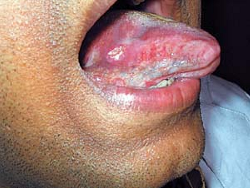 12.3 Squamous cell carcinoma. This persistent, nonhealing ulceration proved to be a squamous cell carcinoma. (Image courtesy of Ashit Marwah, M.D.) |
Erythema Multiforme
See also Chapter 18, “Diseases of Vasculature.”
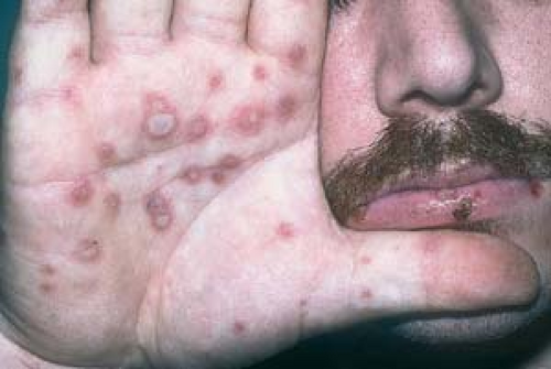 12.4 Erythema multiforme caused by herpes simplex virus. This patient has a recurrent HSV infection. Note the drying crust of the herpetic “cold sore” on his lower lip and the targetlike lesions on his palm. |
Erythema Multiforme Minor
Erythema multiforme minor is a self-limited eruption characterized by symmetrically distributed erythematous macules or papules, which develop into the characteristic targetlike lesions consisting of concentric color changes with a dusky central zone that may become bullous. This is not a disease but a syndrome with multiple underlying causes and associations such as drug reactions, poison ivy contact dermatitis, and, most often, recurrent herpes virus infection.
A crusted lesion of recurrent HSV may be present on the vermilion border of the lip during an outbreak of erythema multiforme (Fig. 12.4).
Erythema Multiforme Major (Stevens-Johnson Syndrome)
This is the more serious variant of erythema multiforme. It has extensive mucous membrane involvement, systemic symptoms, and widespread lesions.
Erythema multiforme major is often accompanied by fever, malaise, myalgias, and severe, painful mucous membrane involvement.
Hemorrhagic crusts on lips and other mucous membranes are seen in addition to the extensive targetoid lesions elsewhere (Fig. 12.5).
Systemic Lupus Erythematosus
Erosions or ulcerations can be seen on the oral mucosa.
See also Chapter 25, “Cutaneous Manifestations of Systemic Disease.”
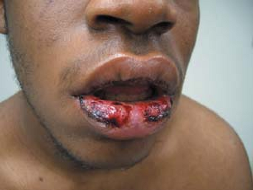 12.5 Erythema multiforme major (Stevens-Johnson Syndrome). Bullae and hemorrhagic crusts are noted on the lips of this patient. He has targetoid lesions elsewhere on his body. |
Perléche
Perléche (derived from the French word meaning “to lick”) is an erythematous eruption that occurs at the corners of the mouth. It is also known as angular cheilitis.
It is sometimes seen in young patients who have atopic dermatitis, specifically atopic cheilitis (Fig. 12.6).
It also appears in the elderly and may be caused by aging and atrophy of the muscles of facial expression that surround the mouth, which results in “pocketing” at the corners of the mouth (Fig. 12.7). These pockets become macerated and serve as nidi for the retention of saliva, resulting in the secondary overgrowth of microorganisms such as yeasts and/or bacteria.
In many instances, perléche is often simply a form of intertrigo, a common inflammatory condition of skin folds that occurs when opposing moist skin surfaces are in constant contact with each other (see Chapter 3, “Psoriasis”).
Other factors such as poor-fitting dentures, malocclusion, anodontia, and bone resorption may lead to drooling or vertical shortening of the face, thus accentuating the melolabial crease.
Lip licking in children, mouth breathing, and orthodontic devices are also risk factors.
Vitamin deficiency is often blamed but rarely proved as a cause of perléche.
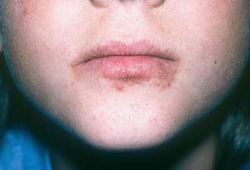 12.6 Perléche (angular cheilitis) and atopic cheilitis. This child has scaling, fissuring, and crusting at the corners of her mouth, as well as eczema of her lips. She also has eczema on other areas of her skin.
Related posts:Stay updated, free articles. Join our Telegram channel
Full access? Get Clinical Tree
 Get Clinical Tree app for offline access

|