Diseases of Vasculature
 URTICARIA AND ANGIOEDEMA
Acute urticaria
Chronic urticaria
Dermatographism
Cold urticaria
Light-induced (solar) urticaria
Cholinergic urticaria
Other physical urticarias
 ERYTHEMA MULTIFORME
 NON-PALPABLE PURPURA AND BENIGN SMALL VESSEL VASCULITIS
Actinic (senile purpura)
Schamberg’s purpura
Majocchi’s purpura (purpura annularis telangiectodes).
HYPERSENSITIVITY VASCULITIS
Palpable purpura
Overview
Cutaneous manifestations of vascular disorders range from a mild rash, urticaria, or angioedema to severe, life threatening vasculitis or anaphylaxis. Diseases featuring inflammation of the wall of blood vessels due to leukocyte migration and resultant damage can affect various organ systems. Large, medium, and/or small vessels may be involved.
Urticaria and Angioedema
Basics
Urticaria, commonly known as hives, is a reaction of cutaneous blood vessels that produces a transient dermal edema consisting of papules or plaques of different shapes and sizes.
Angioedema refers to edema that is deeper than urticaria and involves the dermis and subcutaneous tissue.
By definition, an individual urticarial lesion lasts less than 24 hours.
A total of 10% to 20% of the population has at least one episode of urticaria or angioedema at some point in his or her lifetime.
Pathophysiology
Release of histamine and other compounds by mast cells and basophils causes the appearance of urticaria. Mast cell activation causes degranulation of intracellular vesicles that contain histamine, leukotriene C4, prostaglandin D2, and other chemotactic mediators that recruit eosinophils and neutrophils into the dermis. Histamine and chemokine release lead to extravasation of fluid into the dermis (edema). Histamine effects account for many of the clinical and histologic findings of urticaria.
As with drug reactions, urticaria may be immune mediated or nonimmune mediated (see Chapter 17, “Drug Eruptions”). Causes of urticaria and angioedema include the following:
Immunologic causes, mediated by immunoglobulin E (IgE), include food, drugs, and parasites.
Complement-mediated causes include serum sickness and whole blood transfusions.
Physical stimuli that are non–IgE-mediated include cold, sunlight, and pressure (e.g., dermatographism).
Occult infections include sinusitis, dental abscesses, and tinea pedis. However, such problems are rarely associated with chronic urticaria.
In 85% to 90% of patients with chronic urticaria, the origin is unknown (i.e., chronic idiopathic urticaria).
Classification
Urticaria may be classified as acute or chronic urticaria, physical urticaria, urticarial vasculitis, and hereditary angioedema (rare).
Acute Urticaria
Acute urticaria, by definition, lasts less than 6 weeks.
Outbreaks are often IgE-mediated.
Many patients have an atopic history.
There may be an obvious precipitant such as an acute upper respiratory infection, drug, parasitic infection, or bee sting.
The most common drugs that may cause acute hives are antibiotics (especially penicillin and sulfonamides), pain medications such as aspirin, nonsteroidal anti-inflammatory drugs (NSAIDs), narcotics, radiocontrast dyes, diuretics, and opiates such as codeine.
The most common foods associated with acute urticaria are milk, wheat, eggs, chocolate, shellfish, nuts, fish, and strawberries. Food additives and preservatives such as salicylates and benzoates may also be responsible.
Systemic diseases such as lymphomas and collagen vascular diseases may have an associated urticaria.
Acute urticaria also may be caused by physical stimuli such as pressure, cold, sunlight, or exercise. Such hives are called physical urticarias (see later discussion).
Anaphylaxis or an anaphylactoid reaction can occur.
Chronic Urticaria
Chronic urticaria is, by definition, urticaria that lasts longer than 6 weeks, although the cutoff point is an arbitrary one.
Chronic urticaria occurs in a 2:1 female-to-male ratio.
It is seen predominantly in adults.
The cause is usually unknown; however, chronic urticaria may, very infrequently, be a sign of one of the following systemic diseases: systemic lupus erythematosus (SLE), serum hepatitis, lymphoma, polycythemia, macroglobulinemia, or thyroid disease.
Description of Lesions
Papules and plaques are of varying sizes.
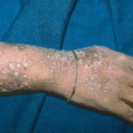
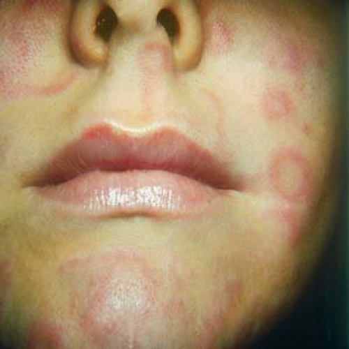
Wheals are the color of the patient’s skin or pale red; a white halo may be noted at the periphery of the lesions (Fig. 18.1).
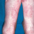
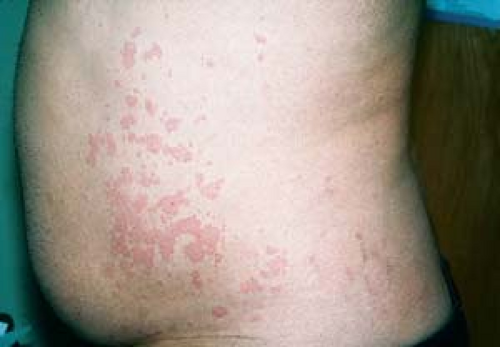
Lesions have various shapes; they can be annular, linear, arciform, or polycyclic, and frequently they are multiple, with bizarre shapes (Figs. 18.2 and 18.3).
Individual lesions disappear within 24 hours (evanescent wheals).
Lesions may be accompanied by a deep swelling (angioedema) around the eyes, lips, and tongue that often looks frightening. Fortunately, angioedema usually lasts less than 24 hours.
 18.1 Urticaria. Skin-colored wheals are present. |
 18.2 Urticaria. Multiple lesions have various shapes and sizes. |
 18.3 Acute urticaria. Lesions are annular, arciform, and polycyclic, with bizarre shapes. |
Distribution of Lesions
Angioedema is noted primarily in the periorbital area (Fig. 18.4), lips, and tongue.
Urticarial lesions may occur anywhere on the body and may be localized or generalized.
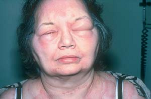 18.4 Urticaria. Marked angioedema is noted. |
Clinical Manifestations
Lesions generally itch.
Arthralgia, fever, malaise, and other symptoms may accompany urticaria when it is the result of hepatitis or serum sickness, for example.
Scratching and rubbing of lesions generally do not produce scabs or crusts similar to those seen in atopic dermatitis.
Episodes of acute urticaria lasts for hours to days (generally less than 30 days).
Approximately 50% of patients with chronic urticaria are free of lesions in 1 year; in other patients, lesions may recur for many years.
Emotional stress may trigger recurrences.
Diagnosis of chronic urtIcaria
The diagnosis of acute and chronic urticarias is usually made on clinical observation and history. If a complete review of systems is normal, and a physical urticaria is ruled out, it is often futile to perform multiple laboratory tests to determine a cause for chronic urticaria. Nonetheless, a positive symptom-directed search for underlying illness (e.g., SLE, thyroid disease, lymphoma, and necrotizing vasculitis) may warrant evaluations such as:
Complete blood count
Erythrocyte sedimentation rate
Fluorescent antinuclear antibody test
Thyroid function studies
Hepatitis-associated antigen test
Assessment of the complement system
Radioallergosorbent test for IgE antibodies
In the presence of eosinophilia, stool examination for ova and parasites
CD203c assay, a new in vitro diagnostic test that is used to detect autoimmune urticaria and identifies antibodies that are responsible for many cases of chronic idiopathic urticaria
Clinical Variants
 Some Physical Urticarias and Their Causes
Dermatographism results from firm stroking or scratching.
Cold urticaria is caused by exposure to cold.
Aquagenic urticaria results from contact with water.
Cholinergic urticaria is caused by heat or exercise.
Solar urticaria results from sun exposure.
Physical Urticarias
Physical factors are the most commonly identified causes of chronic urticaria. Physical urticarias are diagnosed by challenge testing.
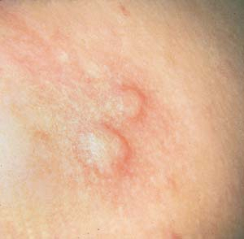
Dermatographism (“Skin Writing”)
Dermatographism affects more than 4% of the general population, in whom it is physiologic and asymptomatic.
Linear erythematous wheals occur 3 to 4 minutes after firmly stroking the skin with the wooden handle of a cotton swab; they fade within 30 minutes. A delayed form also exists.
Lesions are seen under constrictive garments, such as belts and bras, or after a person scratches.
In some persons, itching is the primary symptom.
Episodes of dermatographism may persist for years.
Cold Urticaria
Figures 18.6 A and B.
Cold urticaria occurs mainly in young adults and children.
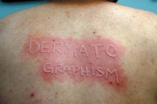
18.5 Dermatographism (“skin writing”). These lesions occurred 3 minutes after stroking with the wooden tip of a cotton swab.
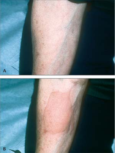
18.6 and B Cold urticaria. The “ice cube test.” Before (A) and 5 minutes after (B) application of an ice cube. A large wheal and surrounding erythema have appeared.
Itchy hives occur at sites of cold exposure, such as areas exposed to cold winds or immersion in cold water.
In the “ice cube test,” a wheal arises on the skin after application of an ice cube.
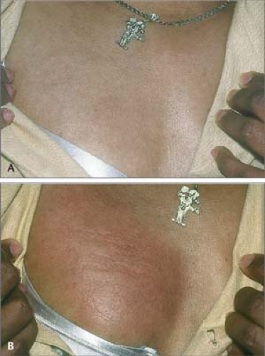 18.7 A and B Solar urticaria induced by ultraviolet A light. Before sun exposure (A) and after 15 minutes of sun exposure through a glass window (B). The glass window filters out ultraviolet B light. |
Light-Induced (Solar) Urticaria
Figures 18.7 A and B.
Solar urticaria occurs in sun-exposed areas of the skin and is triggered by different wavelengths of light.
Cholinergic Urticaria
This type of urticaria is induced by exercise or a hot shower.
The patient exercises to the point of sweating, which provokes lesions and establishes the diagnosis.
Typical lesions are multiple, small, monomorphic wheals.
Other Physical Urticarias
These include those induced by pressure, heat, vibration, and water (aquagenic urticaria).
Insect Bite Reactions
These reactions are also known as papular urticaria. For a full discussion of insect bite reactions, see Chapter 20, “Bites, Stings, and Infestations.”
Reactions to insect bites may be indistinguishable from ordinary hives.
Bites are generally seen on exposed areas.
They may have a central punctum and crust; they also may blister.
Individual lesions may last more than 24 hours.
Erythema Multiforme Minor
See also subsequent detailed discussion.
Lesions are targetoid.
Lesions last more than 24 hours.
Lesions are generally nonpruritic.
Erythema Migrans (Acute Lyme Disease)
For a full discussion of this condition, see Chapter 20, “Bites, Stings, and Infestations.”
Erythema migrans may be indistinguishable from urticaria.
Lesions are usually solitary, annular, and targetlike.
Lesions may last more than 24 hours.
Lesions are generally nonpruritic.
Urticarial Vasculitis
This condition is rare and is probably related to circulating immune complexes.
Persistent hivelike lesions last more than 24 hours.
Lesions may be tender rather than itchy.
Residual purpura or hyperpigmentation often ensues on resolution of lesions.
Evidence of vasculitis (e.g., purpura) is occasionally seen in the lesions.Related posts:

Stay updated, free articles. Join our Telegram channel

Full access? Get Clinical Tree