Fig. 68.1
DLBL-LT: Solitary nodule on the lower leg
Pathology
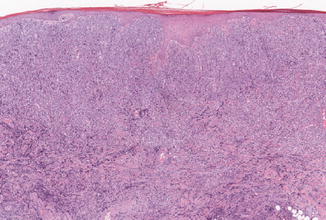
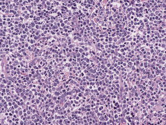
A dense diffuse dermal infiltrate of centroblast- and immunoblast-like cells with non-cleaved, i.e., round nuclei and mitotic activity, is found (Figs. 68.2 and 68.3). There are only a few admixed small lymphocytes or other reactive cells. Rarely, epidermotropic, angiocentric, anaplastic, and spindle cell variants are observed. The tumor cells in DLBL-LT strongly express bcl-2 and MUM-1 as well as IgM, but show weak or no expression of bcl-6 and CD10. Networks of CD21-positive follicular dendritic cells are absent. Deletions in 9p21 (p14(ARF)/p16(INK4a)CDKN2A) are a common finding in DLBL-LT. There is no association with Epstein-Barr virus (EBV).


Fig. 68.2
DLBL-LT: Diffuse dermal infiltrate of blasts

Fig. 68.3
DLBL-LT: Centroblast- and immunoblast-like differentiated tumor cells
Differential Diagnosis
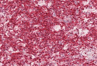
The prognostically most relevant differential diagnosis of DLBL-LT is PCFCL with a diffuse growth pattern. The distinction is based on cytomorphology (centrocyte-like in PCFCL vs. centroblast- and immunoblast-like differentiated tumor cells in DLBL-LT) as well as the phenotype with expression of bcl-2, MUM-1, and FOXP1 in DLBL-LT (Fig. 68.4). Secondary cutaneous involvement by nodal DLBL requires staging. Other blastic neoplasias such as Merkel cell carcinoma, mantle cell lymphoma, or EBV-associated DLBL of the elderly are distinguished by their phenotypic profile and/or EBV status.


Fig. 68.4


DLBL-LT: Characteristic strong expression of bcl-2 by the tumor cells
Stay updated, free articles. Join our Telegram channel

Full access? Get Clinical Tree


