Autoimmune bullous diseases are associated with autoimmunity against structural components that maintain cell-cell and cell-matrix adhesion in the skin and mucous membranes. They include those where the skin blisters at the basement membrane zone and those where the skin blisters within the epidermis (pemphigus vulgaris, pemphigus foliaceus, and other subtypes of pemphigus). The variants of pemphigus are determined according to the level of intraepidermal split formation. There are 5 main variants of pemphigus: pemphigus vulgaris, pemphigus foliaceus, pemphigus erythematosus, drug-induced pemphigus, and paraneoplastic pemphigus. This review focuses only on pemphigus vulgaris.
Clinical presentation of pemphigus vulgaris
Autoimmune bullous diseases are associated with autoimmunity against structural components that maintain cell-cell and cell-matrix adhesion in the skin and mucous membranes. They include those where the skin blisters at the basement membrane zone (bullous pemphigoid, herpes gestationis, mucous membrane pemphigoid, linear immunoglobulin (Ig)A dermatosis, epidermolysis bullosa acquisita, bullous lupus, and dermatitis herpetiformis) and those where the skin blisters within the epidermis (pemphigus vulgaris [PV], pemphigus foliaceus [PF], and other subtypes of pemphigus).
Because of the considerable overlap in the clinical presentation of these conditions, diagnosis of autoimmune bullous skin conditions can be challenging. Detection of tissue-bound and circulating serum autoantibodies and characterization of their molecular specificity is an important modality for diagnosis. In the past decade, there have been several advances in diagnostic modalities for autoimmune bullous skin conditions.
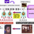
Pemphigus, a word derived from the Greek word “pemphix” meaning bubble or blister, is a life-threatening autoimmune blistering disease characterized by intraepithelial blister formation. Damage to intercellular adhesion structures, desmogleins, are the target of circulating autoantibodies resulting in the hallmark of this condition, acantholysis. Acantholysis may result in the development of the Tzanck phenomenon, which is the rounding of single epidermal cells caused by the loss of cell-cell attachment.
Related posts:
 Diagnosis and Clinical Features of Pemphigus Foliaceus
 Pathogenesis of Endemic Pemphigus Foliaceus
Pemphigoid Gestationis: Pathogenesis and Clinical Features
Pathophysiology of Dermatitis Herpetiformis: A Model for Cutaneous Manifestations of Gastrointestinal Inflammation
Pathogenesis of Endemic Pemphigus Foliaceus
Pemphigoid Gestationis: Pathogenesis and Clinical Features
Pathophysiology of Dermatitis Herpetiformis: A Model for Cutaneous Manifestations of Gastrointestinal Inflammation
 Pathogenesis of Epidermolysis Bullosa Acquisita
Nail Involvement in Autoimmune Bullous Disorders
Pathogenesis of Epidermolysis Bullosa Acquisita
Nail Involvement in Autoimmune Bullous Disorders
Stay updated, free articles. Join our Telegram channel

Full access? Get Clinical Tree


