Autoimmune bullous disorders frequently cause nail abnormalities, particularly paronychia and onychomadesis. In pemphigus vulgaris (PV) nail abnormalities can even precede skin findings. Nail lesions often relapse just before generalized disease exacerbation or recurrence. Severe nail changes are often associated with extensive and severe disease. Fingernails are more commonly affected. A report in the literature associates hemorrhagic nail abnormalities with poor prognosis in patients with PV. Nail scarring and pterygium are a rare complication of bullous pemphigoid. Nail loss has been occasionally reported in epidermolysis bullosa acquisita.
Immunologic characteristics of the nail apparatus
Sinclair and colleagues showed that staining with the epidermal hemidesmosome antigens BP230 kDa, BP180 kDa, and monoclonal antibodies to individual α6 and β4 chains of α6 β4 integrin did not differ in the nail apparatus from normal skin. The lamina lucida and the dermal proteins were also normally expressed in the same distribution as the ubiquitous basal membrane zone (BMZ) antigens, including type IV collagen and laminin 332. All the BMZ antigens and components are also normally expressed in the proximal nail fold, nail matrix, and hyponychium. The desmosomal antigens in this compartment include desmogleins 1 and 3 as in the skin, but the keratin differentiating factors are different.
The normal human nail immune system is very similar to the hair follicle immune system, including a known area of relative “immune privilege” in the proximal nail matrix, which can constitute a safeguard against autoimmunity. Distribution and functional markers of acquired and innate cutaneous immunology differ between the human skin immune system (number and function of antigen-presenting cells are substantially lower in the nail immune system than in the epidermis, with a down-regulation of major histocompatibility complex class II and CD209 expression by Langerhans cells in the proximal nail matrix); natural killer and mast cells are reduced or have diminished function around the human nail apparatus.
Nail involvement in pemphigus vulgaris
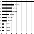
Patients with pemphigus vulgaris (PV) may present with nail abnormalities, which occasionally can precede skin findings. Nail lesions often relapse just before generalized disease exacerbation or recurrence. Severe nail changes are often associated with extensive and severe disease. Nail involvement was previously believed to be rare ; however, the most recent PV prospective study involving 79 patients found that 34.2% had nail changes. Patients with a longer duration of disease and accumulated inflammation are more likely to present with nail changes. Fingernails are more commonly affected, especially the first three, possibly because of more local trauma associated with greater activity of these fingers ( Fig. 1 ). The most common manifestation of PV is paronychia, which can be followed by onychomadesis. However, severe nail dystrophy, discoloration of the nail plate, and even occasionally destruction of the nail plate can occur. A report in the literature associates hemorrhagic nail abnormalities with poor prognosis in patients with PV.
Paronychia
Depending on the location of acantholysis in the nail folds, different nail changes may result.
When the dorsal nail folds are affected, patients present with acute periungual inflammation and bullous lesions, which may be hemorrhagic. When the ventral nail fold is affected, bullae are not visible and the clinical manifestations are indistinguishable from chronic paronychia. The affected nail shows periungual erythema and swelling, with loss of the cuticle, exudation, and crusting around the nail fold. Secondary colonization with bacteria and yeasts may occur.
Onychomadesis/Beau lines
The periungual inflammation can affect matrix keratinization and result in Beau’s lines or nail shedding (onychomadesis). Habibi and colleagues reported these signs in one-third of patients with nail changes caused by PV. Nail matrix damage can also cause nail plate surface abnormalities, such as onychoschizia, cross-ridging, pitting, and trachonychia.
Onycholysis
Subungual bullae cause nail bed detachment with onycholysis, which is often hemorrhagic. Subungual hemorrhages and subungual hyperkeratosis have also been described. In one unusual report, a patient with PV had vegetative verrucous lesions on the periphery of fingernails and digits (mimicking warts) and bullous and vegetating lesions on both feet, with nail plate destruction of two toenails.
Onychomycosis is reported in 25% of patients with PV, with increased prevalence among patients undergoing immunosuppressive therapy.
Diagnosis
The easiest and most painless method of diagnosing PV of the nail is to obtain a biopsy specimen from perilesional skin of the proximal nail fold and perform direct immunofluorescence testing. A positive result will reveal intercellular fluorescence with IgG immunoglobulins and C3. Tzanck tests are difficult to obtain from the ventral nail fold but are very useful in cases of periungual or subungual bullae. Paronychia from PV exacerbation is important to distinguish from other similar clinical presentations, such as acute paronychia from viral (namely herpes simplex infection), bacterial infection, or chronic paronychia. Paronychia in PV has a specific histopathologic feature: suprabasal acantholysis without spongiosis or exocytosis. Bacterial and fungal cultures to exclude superinfection should be performed.
Treatment
Nail PV responds to systemic therapy similarly to other dermatologic manifestations, although it may take longer to resolve. Sometimes, when colonization or superinfection occurs, an associated topical treatment might be helpful.
Nail Disorders and Pemphigoid Group
Nail involvement in bullous pemphigoid is not common and, in most reports, was not confirmed by a nail biopsy. The changes in the nails are thought to be from an immunologic reaction with anti–bullous pemphigoid antigen.
Most commonly bullous pemphigoid affects the nail folds, but the nail bed or matrix can also be involved; the location of the blistering in the different constituents of the nail apparatus will determine the different nail signs (see Fig. 1 ). Paronychia and onychomadesis can occur, as in PV. Nail scarring with atrophy or even permanent loss of the nails has been reported in several cases.
Pterygium of the fingernails has been described in a few patients with cicatricial pemphigoid.
Nail involvement in pemphigus vulgaris
Patients with pemphigus vulgaris (PV) may present with nail abnormalities, which occasionally can precede skin findings. Nail lesions often relapse just before generalized disease exacerbation or recurrence. Severe nail changes are often associated with extensive and severe disease. Nail involvement was previously believed to be rare ; however, the most recent PV prospective study involving 79 patients found that 34.2% had nail changes. Patients with a longer duration of disease and accumulated inflammation are more likely to present with nail changes. Fingernails are more commonly affected, especially the first three, possibly because of more local trauma associated with greater activity of these fingers ( Fig. 1 ). The most common manifestation of PV is paronychia, which can be followed by onychomadesis. However, severe nail dystrophy, discoloration of the nail plate, and even occasionally destruction of the nail plate can occur. A report in the literature associates hemorrhagic nail abnormalities with poor prognosis in patients with PV.
Related posts:
 A Globally Available Internet-Based Patient Survey of Pemphigus Vulgaris: Epidemiology and Disease Characteristics
 Diagnosis and Clinical Features of Pemphigus Foliaceus
Linear IgA Disease: Clinical Presentation, Diagnosis, and Pathogenesis
Pemphigoid Gestationis: Pathogenesis and Clinical Features
Hair Loss in Autoimmune Cutaneous Bullous Disorders
Diagnosis and Clinical Features of Pemphigus Foliaceus
Linear IgA Disease: Clinical Presentation, Diagnosis, and Pathogenesis
Pemphigoid Gestationis: Pathogenesis and Clinical Features
Hair Loss in Autoimmune Cutaneous Bullous Disorders
 Pathogenesis of Epidermolysis Bullosa Acquisita
Stay updated, free articles. Join our Telegram channel

Full access? Get Clinical Tree


