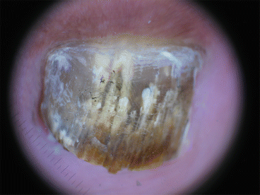
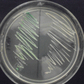
Fig. 13.1
Jagged proximal edge with spikes of the onycholytic area: spiked pattern, indentations at the proximal edge of the area with onycholysis
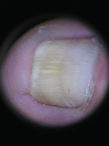
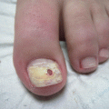
Fig. 13.2
Longitudinal striae. Longitudinal striae of different colors in the onycholytic nail plate

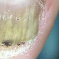
Fig. 13.3
Distal irregular termination or ruin appearance: distal pulverization characteristic of the thickening of the nail plate
In the case of fungal melanonychia, the most frequent dermoscopic finding can be homogeneous pigmentation (brown, gray, or black) which presents as pigmented lines or structureless discoloration (Fig. 13.4); black pigment aggregates can also be seen and can be coarse granules (matte black, roundish structures >0.1 mm) or pigmented clumps (aggregated multiple coarse granules forming larger and irregular-shaped structures) [32].
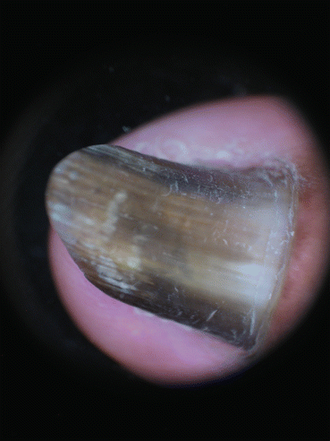
Fig. 13.4
Melanonychia. Homogeneous pigmentation (black) structures and pigmented clumps forming larger and irregular shaped structures with different colors
Other dermoscopic features for differentiating fungal melanonychia from other conditions include [32]:
Multicolored pigmentation (yellow, brown, gray, black or red)
Matte black pigmentation (lines, disrupted black linear pigmentation or homogeneous areas)
Black pigment aggregates (coarse granules or pigment clumps)
Black reverse triangle (wider distally and narrows proximally)
Superficial transverse striation
Blurred appearance
It is important to notice that the coarse granules and pigment clumps correlate with the accumulation of fungal colonies and the pigment produced by the fungi, seen in histopathological samples [32].
Summary for the Clinician
Onychomycosis is one of the most common nail disorders (50 % cases). Accurate diagnosis is important since the treatment can be long-standing and expensive and may be accompanied by severe adverse effects. The diagnosis is made by clinical suspicion along with potassium hydroxide (KOH) examination followed by culture of the sample. This method may be uncomfortable and even painful for the patient and may vary significantly when performed by an experienced mycologist with proper sampling technique. Dermoscopy is a noninvasive tool recently used for the study of nail disorders and helpful to differentiate onychomycosis from traumatic onycholysis or true melanonychia. There are three dermoscopic findings exclusive for onychomycosis: jagged proximal edge with spikes of the onycholytic area, longitudinal striae, and distal irregular termination (ruin appearance). In fungal melanonychia the most frequent dermoscopic findings are homogeneous pigmentation (brown, black, or gray) and black pigment aggregates (coarse granules or pigmented clumps). In fungal longitudinal melanonychia, the band of pigmentation is wider distally and narrows proximally.
Clinical Pearls
Dermoscopy is a noninvasive method that helps to differentiate onychomycosis from traumatic onycholysis or true melanonychia.
There are three dermoscopic findings exclusive for onychomycosis:
- (a)
Jagged proximal edge with spikes of the onycholytic area (characteristic of TDO and DLSO)
- (b)
Longitudinal striae (“aurora borealis pattern” and characteristic of DLSO)
- (c)
Distal irregular termination (ruin appearance) seen in TDO
Fungal melanonychia shows homogeneous or longitudinal pigmentation with black pigment aggregates.
References
1.
3.
4.
Harvey CK, Richardson A. Techniques for obtaining specimens for culture to confirm onychomycosis. J Am Podiatr Med Assoc. 2000;90:394–6.CrossRefPubMed
Related posts:

Stay updated, free articles. Join our Telegram channel

Full access? Get Clinical Tree


