Dermatologic Reactions to Arthropods
James Y.T. Wang
While skin pathology can impact the appearance and health of the skin, external influences also cause important changes in the skin. In this chapter, the focus will be on arthropods that may cause clinically significant disease and irritation of the dermis and epidermis. In many cases, the barrier of the skin is broken via needle-like penetration of arthropod body parts used to access the blood stream. In other cases, arthropod structures release toxins or are themselves toxic to the skin and cause pathology. The chapter is divided into three sections: infestations, bites, and miscellaneous. There are many more diseases caused by arthropods, but the purpose of this chapter is to give a succinct overview of the most commonly encountered conditions caused by arthropods.
 |
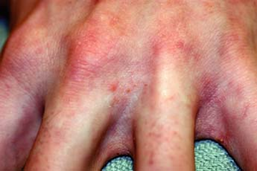
A 20-year-old woman complains of an “itchy rash” on her right arm and hand. Upon examination, it is noted that there are small excoriated papules between her fingers on her right hand and around the right elbow (Fig. 8-1). Five weeks ago, she had gone on a retreat with the volleyball team and everyone had shared bedding. Her roommate is also starting to develop similar symptoms. What is the most likely diagnosis? What test can be done to confirm the diagnosis?
Infestations
Scabies
Background
Scabies is an ectoparasitic skin infestation with the mite Sarcoptes scabiei var. humanus. First described in 1687 with the organism responsible being identified in the 18th century, this intensely pruritic disease was named from the Latin word for “scratch,” scabere. Scabies affect people of all races and age groups and the geographic distribution is worldwide. It is spread by skin-to-skin, via both sexual and nonsexual contact, and in some cases as fomites.
Pathogenesis
The adult mite is 1/3-mm long and has a life cycle of 30 days. An infestation occurs when a fertilized female mite burrows into the stratum corneum of the host’s skin. As it burrows, it lays eggs and expels fecal pellets behind it. A hypersensitivity reaction to the scybala may be responsible for skin irritation and itching because IgE titers are elevated and eosinophilia develop after initial infestation.
Key Features
Hypersensitivity reactions to feces of scabies mite Sarcoptes scabiei are responsible for signs and symptoms.
Pruritic erythematous vesicles, papules, and/or macules are typically distributed in digit web spaces, groin, flexor folds, peri-umbilical regions.
Diagnosis is by skin scrapings and wet mount— visualization of mites, eggs, and/or scybala under microscope.
Treat using topical scabicides, antipruritic medications, and antibiotics (if necessary).
Clinical Presentation
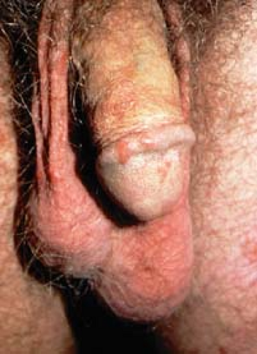
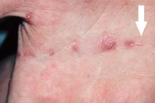
Patients with scabies infestation manifest intractable pruritus, stereotypically with nocturnal exacerbations. Lesions may be eczematous with impetiginization and are often excoriated. Those infected may be asymptomatic for one month following initial infestation by at least one scabies mite. Distribution of skin lesions includes digit web spaces (Fig. 8-1); sides of the fingers; buttocks; peri-umbilical areas; and flexor aspects of the wrists, elbows, and axillary folds. Other areas that may be affected include the penis (Fig. 8-2) and scrotum in men and areolae in women. If noticeable, visualization of a burrow (Fig. 8-3), which can be seen as a short, S-shaped, dark line on the surface of the skin, is pathognomonic for scabies. Family members of patients may also develop similar symptoms and signs. Manifestations of scabies infections are outlined in Table 8-1.
Diagnosis
Differential Diagnosis
Scabies can oftentimes be confused with atopic dermatitis, papular urticaria, pyoderma, insect bites, or dermatitis herpetiformis. Subspecies of nonhuman scabies are also able to cause pruritic eruptions in humans, but these cases do
not have the normal body distribution of human mites and are usually self-limited. Some of these mites include S. scabiei var. canis, S. scabiei var. bovis, Notoedres cati, Cheyletiella yasguri, Cheyletiella blakei, Dermanyssus gallinae, and Ophionyssus natricis.
not have the normal body distribution of human mites and are usually self-limited. Some of these mites include S. scabiei var. canis, S. scabiei var. bovis, Notoedres cati, Cheyletiella yasguri, Cheyletiella blakei, Dermanyssus gallinae, and Ophionyssus natricis.
Table 8-1 Manifestations of Scabies Infection | ||||||||||||||||||
|---|---|---|---|---|---|---|---|---|---|---|---|---|---|---|---|---|---|---|
|
Diagnostic Methods
The visualization of burrows is pathognomonic for scabies, but they may be difficult to find amid excoriations and eczematous plaques. It is generally safe to rely on the characteristic distribution of pruritic lesions seen in scabies. In males, there can also be papules, nodules, and/or ulcers on the penis (Fig. 8-2). A definitive diagnosis requires microscopic identification of mites, eggs, or fecal pellets (scybala). However, in the case that signs of S. scabiei cannot be found but clinical suspicion is high for scabies, it is advisable to proceed with treatment for scabies.
Direct Examination: after applying mineral oil to a skin lesion, scrape or shave area with scalpel blade to remove tops of burrows or papules. Scrapings observed under a microscope under low power should show the scabies mite, eggs, or scybala. Small dermal curettes may also be used to obtain sample. (See Chapter 3, Figs. 3-3 and 3-4 for examples of typical scabies findings seen on scraping.)
Dermoscopy: luminescence microscopy over skin lesions shows small, dark, triangular structures (pigmented section of mite) and a subtle linear segment behind the triangle containing air bubbles (burrow with eggs and fecal pellets).
Potassium Hydroxide Wet Mount: mite feces may dissolve when mount is heated.
Polymerase Chain Reaction (PCR): positive for S. scabiei DNA.
Therapy
Medication is needed to both kill the mites and destroy the eggs. Scabicides must be applied thoroughly to areas behind ears and from the neck down to the soles of the feet. Regions to emphasize coverage are between the fingers and toes, the umbilicus, groin, between buttocks, and under fingernails and toenails. Topical creams ought to be washed off after the recommended time period to avoid toxicity. The morning after topical treatment, all linens, towels, and clothing in the vicinity of the affected individual should be machine-washed and dried using the hot cycles. This ensures that no mites are present to reinfest the host after treatment. Members of the affected household should also be treated with scabicides. Itching and eczema may persist for several months after treatment, even if all mites are successfully removed.
Topical Scabicides
Permethrin 5% cream: apply and leave on overnight. Second application 5 to 7 days later. Not for use in infants younger than 2 months or pregnant/nursing women. Adverse reactions: burning, stinging, pruritus, contact dermatitis (rare).
Lindane 1%: apply and leave on for 8 hours, then wash off. Use for resistant cases only. Not for use in infants, young children, pregnant/nursing women, or patients with seizure disorders or neurologic diseases. Adverse reactions: over-application may lead to central nervous system (CNS) toxicity.
Sulfur (precipitated sulfur, 6%, in petrolatum): apply nightly for 3 nights and wash off thoroughly 24 hours after last application. Not for use in infants younger than 2 months of age, and women who are pregnant or lactating.
Ivermectin (200 mcg/kg × 1 dose, repeat in 7 days): use with caution in elderly patients.
Antipruritic Medications: sedating antihistamines (i.e., hydroxyzine, 25 to 50 mg PO q4h) or short course of topical or systemic glucocorticoids.
Antibiotic Therapy: topical mupirocin or broad-spectrum antibiotics where excoriation has led to impetiginization.
In cases of crusted scabies and scabies in conjunction with human immunodeficiency virus/acquired immunodeficiency syndrome (HIV/AIDS), sequential use of two or more compounds may sometimes be necessary. The entire area of skin should be treated and patients should be isolated until treatment has been completed. The surrounding environment and personal items should be assiduously cleaned.
“At a Glance” Therapy
Topical Scabicides
Permethrin 5% cream: apply and leave on overnight. Second application 5 to 7 days later.
Lindane 1%: apply and leave on for 8 hours, then wash off. Use only for resistant cases.
Sulfur (precipitated sulfur, 6%, in petrolatum): apply nightly for 3 nights and wash off thoroughly 24 hours after last application.
Ivermectin (200 mcg/kg × 1 dose, repeat in 7 days): use with caution in elderly patients.
Antipruritic Medications
Hydroxyzine, 25 to 50 mg PO q4h PRN
Topical corticosteroid: triamcinolone 0.1% BID PRN to AA
Systemic prednisone taper
Antibiotic Therapy: topical mupirocin or broad-spectrum antibiotics such as Keflex 500 mg PO QID × 10 days. Use these only with impetiginization.
Course and Complications
Complications are most often seen in patients with atopic dermatitis. In these individuals, there is an increase in eczematous lesions during the infestation that may persist even after treatment. Excoriations from these lesions can lead to secondary pyoderma and colonization by nephritogenic strains of streptococci, which are most prevalent in tropical regions. The resulting complication is glomerulonephritis. In nonatopics, postscabetic itch can last several months.
Skin lesions do not resolve with scabicide treatment.
Treat close contacts at the same time as the patient. Infections can be passed back and forth.
Launder all bedding and personal items in hot water after treatment.
ICD9 Codes
| 133.0 | Scabies |
Pediculosis
Background
Pediculosis refers to the infestation of certain body areas by head, body, or crab lice. Lice are obligate human ectoparasites that cannot live without their hosts for more than 10 days. The three types of lice, Pediculosis humanis capitis, Pediculosis humanis corporis, and Pthirus pubis, affect different parts of the body, but have similar life cycles and features. Generally, they are 1 to 4 mm long, flat, wingless insects with terminal claws at the end of their three pairs of legs. The legs tend to be clustered toward the anterior aspect of the body, immediately behind the head. The lice live on the hair shafts and crawl down to the
skin to feed on human blood about five times a day. They pierce the skin with their claws, inject anticoagulants and other irritants into the site, and suck blood. Lice are active and travel quickly, resulting in high transmissibility of pediculosis. Most often noticeable in patients with pediculosis is the presence of nits, or eggs, laid by the female lice. Six eggs are laid by the female louse each day for about one month. The nits themselves are less than 1 mm long and are firmly attached to the base of the hair shafts or to fibers on clothing in the case of the body louse. Their proximity to the skin provides nits with the warmth needed to sustain continued growth. Nits are hard to remove, even with a fine-toothed comb.
skin to feed on human blood about five times a day. They pierce the skin with their claws, inject anticoagulants and other irritants into the site, and suck blood. Lice are active and travel quickly, resulting in high transmissibility of pediculosis. Most often noticeable in patients with pediculosis is the presence of nits, or eggs, laid by the female lice. Six eggs are laid by the female louse each day for about one month. The nits themselves are less than 1 mm long and are firmly attached to the base of the hair shafts or to fibers on clothing in the case of the body louse. Their proximity to the skin provides nits with the warmth needed to sustain continued growth. Nits are hard to remove, even with a fine-toothed comb.
Key Features
Lice are bloodsucking insects that reside and lay eggs near the base of hair shafts or in clothing. Each of the three main types of lice has a characteristic distribution on the body (scalp, body, and pubic area).
Presentation is with intense pruritus, excoriations, and secondary impetiginization in affected areas.
Diagnosed by examining plucked hairs for louse or eggs under the microscope.
Treat using topical pediculicides.
Pediculosis capitis
A worldwide infestation caused by bloodsucking, wingless insects, Pediculus humanus capitis, who live on head hair and feed on blood from the scalp.
Diagnosis confirmed by presence of nits attached to head hairs or identification of a louse.
Head lice is spread by head-to-head contact and fomites.
Worldwide resistance to traditional treatments (pyrethrin, Permethrin) is increasing.
Pediculosis corporis
Body lice is caused by an infestation of humans and their clothing by Pediculus humanus corporis.
Body lice lay their eggs in clothing, not on people.
Commonly found in those in crowded, unsanitary living conditions.
Lice may transmit epidemic typhus, trench fever, and relapsing fever.
Pediculosis pubis
May involve pubic as well as other hair-bearing sites such as the beard, eyelashes, axillae, and perianal regions.
Transmission via sexual, close contact, or rarely through contaminated clothing, towels, and bedding.
Clinical Presentation
The bite of the louse itself is painless, but hypersensitivity to the saliva and feces of the mite may lead to inflammation and pruritus. Immediate urticarial lesions may appear in some individuals, but most develop small macules or papules hours to days later. Intense scratching of these areas can result in trauma to the skin, excoriations, and secondary bacterial infections. Most often seen are the results of pruritus. A comparison of the different types of pediculosis is presented in Table 8-2.
Pathogenesis
Saliva and fecal material can induce a hypersensitivity reaction and inflammation. The resulting pruritus leads to scratching and the development of excoriations and secondary impetiginization of the affected area(s).
Pediculosis Capitis
There are over 12 million cases per year of head lice in the United States alone. The area of infestation is limited to the human head and is commonly seen on the back of the head and neck and behind the ears. It occurs primarily in children, especially in girls. African Americans have very low incidence of head lice. Head lice are spread by close physical contact and sharing of head gear, combs, brushes, and pillows. Usually, an infestation on an individual will have less than 20 adult lice.
Clinical Features
Intense pruritus leads to scratching, resulting in inflammation and secondary bacterial infection. Common clinical features include excoriations with pustules, crusting, and maculae ceruleae (blue to slate-gray macule) in the head and neck region as well as conjunctivitis and cervical lymphadenopathy.
Table 8-2 Comparison of Head, Body, and Crab Lice | ||||||||||||
|---|---|---|---|---|---|---|---|---|---|---|---|---|
|
Diagnosis
Differential diagnosis.
Conditions that resemble head lice include the presence of seborrheic scales, hair casts, and hairspray.
Diagnostic methods.
Live nits on proximal head hair shafts can often be seen with the naked eye in cases of Pediculosis capitis infestation. Examine plucked hairs under the microscope to determine whether the nits contain live larvae. The presence of live adult lice, immature nymphs, or viable eggs confirms the diagnosis.
Pediculosis Corporis
The structure of body lice looks similar to head lice, but body lice tend to be larger. It is an infestation associated with poverty and poor living condition. Therefore, it is most often seen in the poor, homeless, recent refugees, and soldiers in wartime conditions. The main mode of transmission is via contaminated bedding or clothing because body lice do not lay eggs on humans, but rather in clothes that the person wears.
Clinical Presentation
The presence of maculae ceruleae in areas where clothing most commonly contacts a person, such as the waistband and on buttocks and thighs, is highly suspicious for body lice. The lesions may or may not be pruritic, but there will be linear excoriations, sometimes with secondary impetiginization, seen on the trunk from the intense pruritus. Adult lice are rarely seen.
Diagnosis
Diagnostic methods.
Careful examination of clothing for nits, especially in the seams, is crucial. An effective method may involve shaking out the patient’s clothing over a sheet of newspaper. Lice may then be seen moving on the paper.
Associated diseases.
Body lice are vectors for Rickettsia prowazekii (epidemic typhus); Rickettsia quintana (trench fever); and Borrelia recurrentis (relapsing fever).
Pediculosis Pubis
The crab louse (Phthirus pubis) is physically distinctive from head and body lice. Its body is more rounded, resembling the shape of a crab, and has progressively larger claws from its front pairs of legs toward the rear pairs. This formation allows the louse to grip sparser, coarser hairs of the pubic area, eyebrows, eyelashes, thighs, and perianal regions. The incidence of crab lice is slightly higher in men, potentially due to larger amounts of coarse body hair. It is considered a sexually transmitted disease and is associated with other sexually transmitted infections. There are no racial differences in incidence of infestation, although pediculosis pubis often recurs in men who have sex with men (MSM).
Clinical Presentation
Maculae ceruleae are often present in the pubic region. Patients will present with intense itching and excoriations in the infested areas. Local lymphadenitis and
fever may present concurrently due to secondary bacterial infection of those excoriations. Hairier individuals may also have infestations of hairs of the thighs, trunk, and perianal area. Even beard, moustache, and eyelashes can be affected. Infestation of eyelashes and periphery of scalp occurs mainly in children and may indicate sexual abuse. Extra-pubic infestations, such as those of the beard, moustache, and eyelashes are more common in the homeless population.
fever may present concurrently due to secondary bacterial infection of those excoriations. Hairier individuals may also have infestations of hairs of the thighs, trunk, and perianal area. Even beard, moustache, and eyelashes can be affected. Infestation of eyelashes and periphery of scalp occurs mainly in children and may indicate sexual abuse. Extra-pubic infestations, such as those of the beard, moustache, and eyelashes are more common in the homeless population.
Diagnosis
Diagnostic methods.
Examine plucked hairs under microscopy for louse or nits.
Therapy
Pediculosis Capitis
Topical pediculicides
Synergized pyrethrin creams (RID, A-200, R&C shampoo)
Permethrin 5% cream: hair should be combed with fine-toothed comb to remove remaining nits
Malathion lotion 0.5% (Ovid): kills all lice and ova in 10 minutes
Lindane (gamma-benzene hexachloride) 1% shampoo
Benzyl alcohol 5% lotion
Environmental control
Use fine-toothed comb to remove as many nits as possible. An 8% formic acid rinse helps to dislodge nits from their attachment to the hair.
Members of household should be examined. Treatment of household members up to discretion of physician.
Following therapy, clothing and bedding should be washed and dried using hot cycle; nonwashables should be dry cleaned, ironed, or put in the clothes dryer without washing. Wash combs and brushes in very hot water, can add pediculicides first.
“No nit” policies excluding children with nits from attending school are excessive.
Pediculosis Corporis
Discarding or laundering infested clothing and restoring proper hygiene should cure the infestation because body lice lay eggs in the clothing and not on the human body.
Bedding should be laundered with hot water, boiled, or discarded.
Consider treating the patient head to toe with a single application of Permethrin 5% cream.
Pediculosis Pubis
Use topical treatments (synergized pyrethrins) as in cases of Pediculosis capitis, but in areas affected by crab lice specifically, such as the pubic and perianal regions. A common cause of treatment failure is neglect of thighs, trunk, and axillary regions in hairier individuals. Sexual contacts should be treated simultaneously and clothing/bedding should be washed and dried using the hot cycle.
Parasitophobia is often a consequence postinfestation.
Specific agents: therapy for eyelash involvement is applying petrolatum twice daily for 8 days, followed by removal of nits. HIV/AIDS patients may be unresponsive to conventional therapy.
There are signs of local resistance to topical medications for treatment of pediculosis. In resistant cases, systemic therapy with ivermectin (adult dosage [for >5 years; safety in <5 not established]: 150 to 200 mcg/kg/d PO as single dose; 12 mg PO repeated in 7 to 10 days) is indicated.
“At a Glance” Therapy
Pediculosis Capitis
Topical pediculicides
First-line:
Synergized pyrethrin creams (RID, A-200, R&C shampoo)
Permethrin 5% cream
Second-line:
Malathion lotion 0.5% (Ovid): kills all lice and ova in 10 minutes.
Lindane (gamma-benzene hexachloride) 1% shampoo
Benzyl alcohol 5% lotion
Environmental control
Use fine-toothed comb to remove as many nits as possible.
8% formic acid rinse helps to dislodge nits from their attachment to the hair.
Examine and treat close contacts.
Following therapy, clean bedding and local environment.
Pediculosis Corporis
Discard/launder infested clothing and restoring proper hygiene should cure the infestation.
Bedding should be laundered with hot water, boiled, or discarded.
Consider treating with a single application of Permethrin 5% cream.
Pediculosis Pubis
Use topical treatments (synergized pyrethrins) as in cases of Pediculosis capitis.
Sexual contacts should be treated simultaneously.
Clothing/bedding should be washed and dried using the hot cycle.
Eyelash involvement: petrolatum twice daily for 8 days, followed by removal of nits.
Resistant infections
Systemic therapy with ivermectin
Adult dosage (for >5 years): 150 to 200 mcg/kg/d PO as single dose; 12 mg PO repeated in 7 to 10 d)
Safety in children younger than 5 years not established
Refer to a dermatologist when topical therapies fail to eradicate the infestation.
Associated disease that may be transmitted especially by body lice (epidemic typhus, trench fever, and recurrent fever).
Consider medication resistance in recalcitrant cases.
ICD9 Codes
| 132.0 | Pediculus capitis (head louse) |
| 132.1 | Pediculus corporis (body louse) |
| 132.2 | Phthirus pubis (pubic louse) |
| 132.3 | Mixed pediculosis infestation |
| 132.9 | Pediculosis, unspecified |
Bites
Key Features
Spiders are carnivorous arachnoids that have fangs containing venom.
Most bites occur when spider is pressed against skin or threatened.
The bites of most spiders that are venomous lead to mild localized reactions around the area of the bites.
Treatment for most spider bites include rest, ice, and elevation of the affected area.
Some spider bites are more serious and may require more aggressive treatment.
Spiders
Spiders are carnivorous arthropods with four sets of legs. They are generally not aggressive and bite humans only when threatened. They have fangs containing venom, which are usually used to catch and immobilize prey. Spider venom usually contains inflammatory factors that simply cause pain and swelling. However, some may have neuromuscular effects and can lead to significant necrosis of the skin and underlying tissues. Although some spider bites may not be felt at the time of the initial bite, most do cause pain at the instant they occur. Localized pain, swelling, itching, erythema, blisters, and necrosis may be found, along with two puncta (fang marks) in the center. The swelling
appears at the bite site, expanding radially to a few centimeters, and can be significant for some species of spiders.
appears at the bite site, expanding radially to a few centimeters, and can be significant for some species of spiders.
For most spider bites, the lesions resolve spontaneously. Treatment of itching and swelling is sufficient by applying ice and taking antihistamines. However, some spider bites are more venomous and may require further treatments.
Most spiders in the United States are not able to bite humans due to small mouth parts. There are two spiders that are able to bite and envenomate humans, the black widow (Lactrodectus spp.) and the brown recluse (Loxoscelidae reclusus) spiders, which will be examined in more detail. There are other spiders which may bite humans, but these are even rarer than these examined in this section.
Black Widow Spider
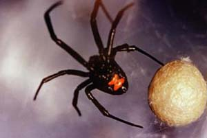 |
A 35-year-old man is re-stacking an old pile of wood that had fallen over during winter. After picking up a piece of firewood, he feels an acute pain on the dorsum of his right hand. He sees a black shiny spider on the ground nearby that had obviously fallen from the piece of wood (Fig. 8-4). His hand is swelling and two punctae are seen. What should he do next?
Stay updated, free articles. Join our Telegram channel

Full access? Get Clinical Tree


