Fig. 32.1
Dermatofibrosarcoma protuberans. A typical presentation characterized by a slow-growing multinodular indurated plaque on the anterior trunk of a 30-year-old woman
Pathology
DFSP is characterized by a dense proliferation of uniform spindle cells arranged into a distinctive, monotonous storiform pattern that occupies the entire dermis and extends into the septa and lobules of subcutaneous fat in a characteristic “honeycomb” pattern (Figs. 32.2, 32.3, and 32.4). The spindled cells that have monomorphous elongated nuclei and scanty pale cytoplasm are characteristically arranged in a “cartwheel” pattern (Fig. 32.5). Mitotic activity is scanty and does not exceed five mitoses per 10 high-power fields. Foci of myxoid degeneration may be present and the neoplasm exhibits only minimal, if any, necrosis. Apart from the conventional variant, DFSP can present with various histological patterns. Histological variants of DFSP include: giant cell fibroblastoma and pigmented, myxoid, myoid, atrophic, sclerotic, granular cell, and fibrosarcomatous DFSP. Giant cell fibroblastoma, a variant of DFSP described only in children, is composed of spindle cells usually arranged in a storiform pattern but frequently with areas of myxoid stroma and distinctive pseudovascular spaces lined by multinucleated giant cells. Pigmented DFSP, or Bednar tumor, has melanin-containing dendritic cells in variable concentrations among the spindle cells arranged in the typical storiform pattern. Myxoid DFSP exhibits prominent myxoid stromal changes that can efface the characteristic storiform pattern, a multinodular growth pattern, and a prominent capillary vasculature. Atrophic DFSP is less cellular than classic DFSP and is characterized by a marked atrophy of the middle dermis. Fibrosarcomatous DFSP exhibits areas of classic low-grade DFSP interspersed with more cellular areas where the cells are arranged in fascicles disposed in a herringbone pattern. These areas manifest significant cellular atypia and increased mitotic activity. Very rare cases of fibrosarcomatous DFSP with giant rosettes and DFSP with unusual sarcomatous transformation, which mimic undifferentiated pleomorphic sarcoma/malignant fibrous histiocytoma, have been reported. Exceptional cases of purely subcutaneous DFSP, histologically indistinguishable from typical DFSP, have been described. Areas resembling neurofibroma or schwannoma with nuclear palisading and even Verocay body formation can sometimes be encountered.
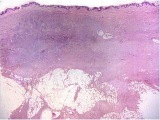
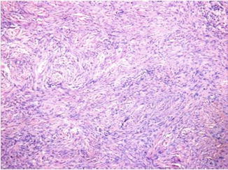
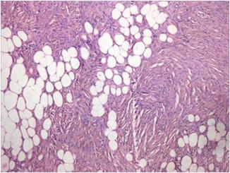
Fig. 32.2
The neoplasm occupies the entire dermis and extends into the septa and lobules of subcutaneous fat all the way down to the fascia
Fig. 32.3
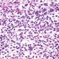
DFSP is characterized by a dense proliferation of uniform spindle cells arranged into a distinctive, monotonous storiform or “cartwheel” pattern
Fig. 32.4


The spindle cell proliferation extends into the septa and lobules of subcutaneous fat in a characteristic “honeycomb” pattern
Related posts:


Stay updated, free articles. Join our Telegram channel
Full access? Get Clinical Tree
