Dermatitis
Gunilla Carlsson Thorn
ATOPIC DERMATITIS/ECZEMA
I. BACKGROUND
Atopic dermatitis (AD) is an intensely pruritic, chronic skin disease that can affect all age groups, but usually arises within the first 5 years of life. AD can be classified into three stages: infantile, childhood, and adulthood. The infantile form tends to be highly inflammatory and favors the face and the extensor surfaces. The adulthood form tends to show low-grade inflammation and marked lichenification and favors the flexural surfaces. The childhood form can show features of both the infantile and the adulthood form, but with time will progress to favor the adulthood form.
AD is a common condition with an estimated prevalence of 10% to 20% in American schoolchildren. Increased frequencies of AD are seen in individuals with parental history of atopy; however, the environment also seems to play a role in the development of AD. Specifically, individuals within highly industrialized societies and advantaged socioeconomic classes are at an increased risk for developing AD. The pathogenesis of AD is highly multifactorial and not completely understood. It is generally agreed, however, that AD is related to immunologic abnormalities, compromised skin barrier function, blood vessel reactivity, and abnormalities in cutaneous nerves and neuropeptides. Notable immunologic abnormalities include activation of the T-helper 2 response, elevated serum immunoglobulin E levels, peripheral blood eosinophilia, and abnormally exuberant immune reactions to various antigens including allergens and microbial agents. Individuals with AD not only have exuberant reactions to microbial agents but also have increased susceptibility to microbial agents. It is estimated that over 90% of AD lesions are colonized with microbial agents, most commonly with Staphylococcus aureus (S. aureus). This increased susceptibility is attributed not only to the compromised skin barrier but also to the reduced expression of antimicrobial peptides seen in AD.
II. CLINICAL PRESENTATION
All forms of AD share the feature of intense pruritus and xerosis in a relapsing and remitting course. The pruritus is often worse in the evening and can be exacerbated by heat, sweating, and contact with wool clothing. It is the scratching and rubbing resulting from this pruritus, which typically initiates the formation of the classic AD skin lesions. Xerosis is seen in the majority of individuals with AD and generally involves the entire skin surface, not only areas affected by dermatitis.
The infantile form usually presents in the first 2 to 12 months of life and is characterized by intensely pruritic, brightly erythematous papules and plaques. These lesions often have overlying vesicles, oozing, serous crusting, and secondary excoriations and are distributed over the face (especially the cheeks), scalp, and extensor surfaces (Fig. 9-1). The diaper area is classically spared.
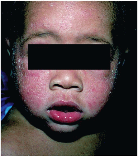 Figure 9-1. Atopic dermatitis, infantile form. (From Goodheart HP. Goodheart’s Photoguide of Common Skin Disorders. 2nd ed. Philadelphia, PA: Lippincott Williams & Wilkins; 2003.) |
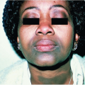
The childhood form defines AD in individuals between 2 and 12 years of age. The clinical features usually resemble the infantile form early on and over time evolve toward the adulthood form. The adulthood form is characterized by subacute to chronic lesions with increased scale and lichenification and decreased erythema, vesiculation, oozing, and crusting (Fig. 9-2). The lesions favor the flexural areas including the neck, antecubital fossa, and popliteal fossa, but can also commonly involve the hands and eyelids. Additional features commonly associated with AD are listed in Table 9-1.
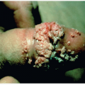
The most common complication seen in the setting of AD is secondary infection. The most common agent causing secondary infection is S. aureus. It should be suspected when the AD lesions show prominent crusting and oozing. Treatment with antistaphylococcal antibiotics with good skin penetration is then indicated. Patients with AD are also predisposed to cutaneous viral infections, including warts, molluscum contagiosum, and herpes simplex virus (HSV). Infection of AD lesions with HSV presents with numerous monomorphic punched-out erosions or hemorrhagic crusts known as eczema herpeticum (Fig. 9-3).
III. WORKUP
AD is a clinical diagnosis and typically no additional workup is needed. Features that support a diagnosis of AD include pruritus, involvement of flexural surfaces in children and adults, involvement of face and extensor
surfaces in infants, a personal or family history of asthma, seasonal allergies or AD, and a history of dry or sensitive skin. If the presentation is atypical, a skin biopsy can be performed; however, it is important to be aware that all forms of dermatitis, regardless of the cause, will show similar histologic features. Although questions regarding food allergens are often raised by patients or their
families, food allergens are rarely associated with AD and such allergy testing is generally not advantageous. If there is a concern for secondary infection, appropriate cultures should be obtained.
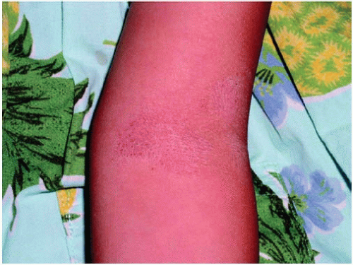 Figure 9-2. Atopic dermatitis, adult form. Note the flexural and hand involvement. (Courtesy of Esther K. Chung, MD.) |
TABLE 9-1 Clinical Features Associated with Atopic Dermatitis | ||||||||||||||
|---|---|---|---|---|---|---|---|---|---|---|---|---|---|---|
|
Related posts:

Stay updated, free articles. Join our Telegram channel

Full access? Get Clinical Tree


