Deceased Donor Transplantation Hepatectomy (Artery, Portal Vein, and Bile Duct)
Karim J. Halazun
Gerardo Kahane
James V. Guarrera
DEFINITION
Removing the recipient liver can be one of the most challenging parts of the liver transplant. Care must be taken to assure that the hepatectomy is done safely with minimal blood loss in order to avoid major complications. Dissection of the hilum and isolation of the vessels and duct is an integral part of the liver transplant procedure and needs to be carried out with care. This chapter will describe our approach to the recipient hepatectomy with particular attention to the hilar dissection.
IMAGING
Whenever possible and where available, cross-sectional imaging of the patient should be reviewed preoperatively, with particular attention being paid to the patency of the portal vein, presence of any vascular anatomic anomalies, presence of large hilar collateral vessels, and presence of large spontaneous portosystemic shunts, which may require ligation to augment portal flow.
PATIENT POSITIONING
The patient is placed in the supine position with the right arm tucked. We routinely use a Thompson retractor for the liver transplant procedure.
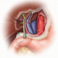
Various incisions have been adopted and used for liver transplantation. We use a chevron incision with extension to the right side all the way to the flank. The left side of the incision is taken to the edge of the rectus muscle. This allows adequate access to the right retroperitoneum and the left lobe of the liver. Wide exposure of the upper abdomen at the costal margin facilitates dissections of the hepatic veins and suprahepatic inferior vena cava (IVC). Excellent exposure of these areas is critical to facilitate clamping and allograft outflow reconstruction.
TECHNIQUES
SUMMARY OF MOBILIZATION OF THE LIVER AND INFERIOR VENA CAVA
Take down the falciform ligament to expose the anterior hepatic veins.
Once the anterior portion of all three hepatic veins is exposed, turn your attention to the left triangular ligament.
The surgeon should place their right hand or the edge of a laparotomy pad behind the ligament to protect the stomach and spleen and cauterize directly onto their finger (or the pad).
Occasionally, the triangular ligament is deep and the left lobe extends beyond the spleen. In these cases, place a laparotomy pad behind the left lobe under the left triangular ligament and use this to protect the posterior structures. Avoid excess traction on the tip of the left lateral segment.
Dissect the triangular ligament to the left hepatic vein. In most cases, there is a left phrenic vein that requires ligation.
Rotate the patient to the right, and lift the left lobe superiorly and to the right to expose the lesser omentum (pars flaccida) overlying the caudate lobe.
Incise the clear space overlying the caudate using cautery and tie as needed toward the left hepatic vein.
There is usually a band of tissue that lies immediately between the caudate lobe and left hepatic vein that will need to be ligated to allow full access to the posterior aspect of the left hepatic vein.
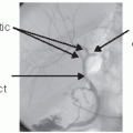
A replaced left hepatic artery from the left gastric artery is found in the lesser omentum and will need to be securely ligated during this part of the dissection.
Turn the patient back to the left and place them in reverse Trendelenburg position. This will aid the dissection and mobilization of the right lobe.
Expose the anterior right hepatic vein. An avascular tissue plane exists between the liver and diaphragm. In this plane, dissect the diaphragm off of the liver.
Once completed, lift the right lobe superior and medially, exposing the retroperitoneal attachments of the liver to the right kidney and adrenal gland. Dissect these attachments with cautery and expose the IVC inferiorly.
Dissect the areolar tissue anterior to the infrahepatic IVC, exposing the lateral edge of the IVC. Once exposed, the medial edge can also be exposed by dissection of the tissue between the IVC and portal vein.
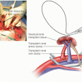
Pass a right-angle clamp under the IVC and encircle with an umbilical tape. Some surgeons do not encircle the IVC, especially if not doing a bicaval reconstruction.
Once the infrahepatic cava is encircled, free the posterior part of the IVC from the retroperitoneum, continuing the dissection along the left side, allowing the IVC to be lifted off of the retroperitoneum.
Further retrocaval dissection from the right side allows full exposure of the right hepatic vein. Pass your finger behind the cava from the right to the left side, ensuring the completion of the caval dissection for a bicaval liver transplant.
The mobilization of the liver and IVC are now complete, and the hilar dissection (if not already performed) should now be undertaken.
Some important variations in the caval dissection for the caval sparing technique:
If a piggyback or cavocavostomy approach is to be undertaken (both require caval sparing) as well as
use of a partial allograft, the IVC does not need to be lifted off of the retroperitoneum.
The caval sparing approach requires ligating the hepatic veins. This is best done with an endovascular stapler. Also ligate the short hepatic veins entering the cava from the caudate lobe of the liver, starting inferiorly and moving up to the hepatic veins. This is best done after the hepatic arteries are ligated because it minimizes bleeding from the liver.
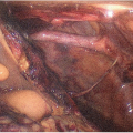
THE HILAR DISSECTION
The hilum of the liver can be approached early in the case or after mobilization of the liver. The decision to approach the hilum early or late depends onRelated posts:

Stay updated, free articles. Join our Telegram channel

Full access? Get Clinical Tree