Fig. 74.1
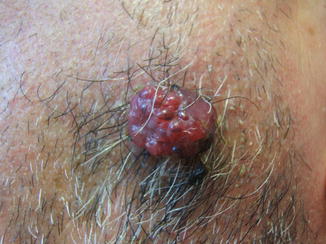
Cutaneous metastases from clear cell carcinoma of the kidney. The most common clinical presentation is characterized by asymptomatic, pink to red to purplish, angiomatous subcutaneous nodules mimicking vascular lesions

Fig. 74.2
Cutaneous metastases from clear cell carcinoma of the kidney. Close-up of angiomatoid skin metastasis
Pathology
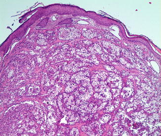
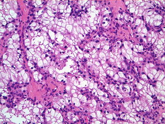
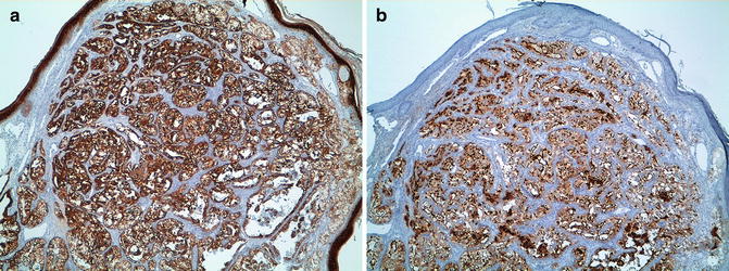
Almost all cases are of clear cell histological type with tumor cells containing abundant glycogen and some lipids. There is often a grenz zone with uniform polyhedral, atypical clear cells arranged in compact, trabecular, tubulocystic, alveolar, or papillary configurations (Figs. 74.3 and 74.4). Prominent vessels and red blood cell extravasations with hemosiderin deposits in the stroma are common features. Neoplastic cells are positive for vimentin, cytokeratins AE1/AE3 (Fig. 74.5a), and MNF116. CD10, the common acute lymphoblastic leukemia antigen, is expressed in 89–100 % of renal cell carcinoma (Fig. 74.5b) and is a useful marker to distinguish cutaneous metastatic renal cell carcinoma from primary cutaneous adnexal carcinomas with eccrine and apocrine differentiation. Renal cell carcinoma marker (RCC-Ma) is another marker which is expressed in 79.7 % of primary renal cell carcinoma (96 % of papillary variants) and whose expression is highly specific (98 %) for metastatic renal cell carcinoma. PAX2 and especially PAX8 are diagnostically useful markers for both primary and metastatic renal neoplasms of different histological types.


Fig. 74.3
Cutaneous metastases from clear cell carcinoma of the kidney. Under a grenz zone, there are uniform polyhedral, atypical clear cells arranged in trabecular and alveolar configurations

Fig. 74.4
Cutaneous metastases from clear cell carcinoma of the kidney. Clear cell histological type with tumor cells containing abundant glycogen

Fig. 74.5
Cutaneous metastases from clear cell carcinoma of the kidney. (a) Neoplastic cells are positive for cytokeratins AE1/AE3 (b) and CD10
Differential Diagnosis
Because of the prominent vascular supply of renal cell carcinoma, cutaneous metastases are known to have an angiomatous appearance that mimics pyogenic granuloma, hemangioma, Kaposi sarcoma, and Kaposi sarcoma. It is important to consider skin metastasis in case of new onset tumors with a vascular appearance in the head and neck region of patients with associated renal malignancy. Histologically, all clear cell tumors of the skin enter the differential diagnosis. RCC-Ma is an immunohistochemical marker that is mostly expressed by renal cell carcinoma and is negative in other clear cell tumors of the skin. Adipophilin, a marker expressed in neoplasm with sebaceous differentiation, is also positive in cutaneous metastases from renal cell cancer.
Prognosis
Metastatic cutaneous renal cell carcinoma is generally regarded as a late manifestation of the disease leading to a poor prognosis with a median overall survival rate of approximately 6–9 months. Clinicopathologic parameters of the primary renal cell carcinoma that are associated with a better prognosis include size less than 5 cm in diameter; lack of diffusion to the collecting system, perirenal fat, or regional lymph nodes; and a predominance of clear cells.
Treatment
Chemotherapeutic treatments are unsatisfactory, with maximal partial response rates of 5–20 % reported. The use of new therapeutic agents directed towards aberrant molecular pathways such as tyrosine kinase inhibitors (sorafenib, sunitinib) or antiangiogenic substances, such as VEGF receptor antagonists, has shown antitumor activity and clinical benefit. For palliative treatment of skin metastases, surgical excision of solitary skin lesions, radiotherapy alone or combined with surgery, or intralesional injection of therapeutic agents such as interferon has been used with variable results. Curiously, in the appropriate setting, surgical excision of isolated cutaneous metastases using Mohs surgery has been proposed.
Stay updated, free articles. Join our Telegram channel

Full access? Get Clinical Tree


