Fig. 73.1
Multiple isolated nodules over the neck and chest area from a lady with known lung carcinoma

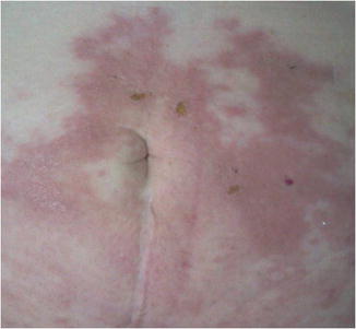
Fig. 73.2
Cutaneous metastases of lung carcinoma. Skin metastases can rarely present as inflammatory carcinoma

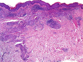
Fig. 73.3
Cutaneous metastases of lung carcinoma. Tumoral deposits of lung adenocarcinoma in the dermis accompanied by an abundant inflammatory infiltrate

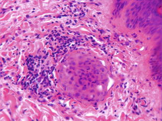
Fig. 73.4
Cutaneous metastases of lung carcinoma. Typical inflammatory carcinoma with tumoral deposits in dilated lymphatics
Pathology
Histologically, undifferentiated lung cancer (Fig. 73.3) is the type that most frequently metastasizes to the skin (50 %), followed by adenocarcinoma and squamous cell carcinoma (30 % each). The following histological patterns of pulmonary cutaneous metastases are usually seen: undifferentiated, adenocarcinoma, and squamous cell carcinoma. Much less commonly, bronchiolar carcinoma, mucoepidermoid carcinoma, carcinoid, pulmonary sarcoma, small cell carcinoma, or the rare intravascular bronchioalveolar tumor may be seen. Cutaneous metastases may also display signet-ring cells and an adenoid cystic carcinoma-like pattern.
Immunohistochemistry: Metastases from lung adenocarcinoma display the following immunoprofile: CK7+, CK20 usually negative, CAM5.2+, CEA+, TTF1+, and BerEP4+. Metastatic cutaneous squamous cell carcinoma of the lung is positive for CK5/6, CK 19, and CEA and negative for CK7, CK20, CAM5.2, TTF1, and BerEP4. Metastatic small cell carcinoma of the lung stains with low-molecular-weight cytokeratin in a diffuse perinuclear pattern and variably with antibodies against neurofilament.
Stay updated, free articles. Join our Telegram channel

Full access? Get Clinical Tree


