Fig. 71.1
Cutaneous metastases of breast carcinoma. Warm, erythematous patches and plaques surrounding the sectorectomy scar for breast carcinoma

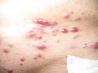
Fig. 71.2
Cutaneous metastases of breast carcinoma. Multiple tumors with a vascular appearance disseminated on chest and abdomen in a patient with breast cancer

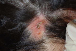
Fig. 71.3
Cutaneous metastases of breast carcinoma-alopecia neoplastica. Erythematous indurated alopecic plaque on the scalp of a patient with breast carcinoma
Pathology
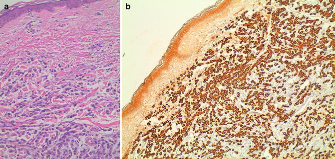
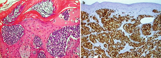
In general, the histological features of the metastases are similar to those of the primary tumor, although metastases may exhibit less differentiation. Histological variants include a glandular pattern, an Indian file pattern of malignant cells in between collagen fibers (Fig. 71.4a, b), lymphatic embolization by malignant cells, a fibrotic pattern, and an epidermotropic pattern (Fig. 71.5a, b). Inflammatory metastatic carcinoma shows deposition of malignant cells in superficial and/or deep lymphatics accompanied by a slight inflammatory infiltrate. Telangiectatic carcinoma displays aggregations of malignant cells in both lymphatics and blood vessels and many dilated blood vessels in the papillary dermis. Sclerodermoid metastatic lesions present with tumoral deposits accompanied by a fibrotic stroma. Carcinoma en cuirasse shows prominent fibrosis within which the few tumor cells usually encountered can be easily overlooked. Nodular metastases of breast carcinoma tend to display masses of tumor cells (Figs. 71.6 and 71.7), although some may also show a linear distribution of tumor cells in between collagen bundles. In alopecia neoplastica, the pilosebaceous units are destroyed by either neoplastic cells or fibroplasia.


Fig. 71.4
Cutaneous metastases of breast carcinoma. (a) Indian file pattern. (b) Positivity of tumor cells for cytokeratin AE1/AE3

Fig. 71.5


Cutaneous metastases of breast carcinoma. (a) Epidermotropic metastasis. (b) Tumor cells are estrogen receptor positive
Stay updated, free articles. Join our Telegram channel

Full access? Get Clinical Tree


