Cutaneous Manifestations of HIV Infection
Mary Ruth Buchness
Overview
The first organ that may be affected in human immunodeficiency virus (HIV) infection is the skin. Before the advent of highly active antiretroviral therapy (HAART), the inevitable decrease in CD4 cells with disease progression was accompanied by a variety of HIV-associated skin diseases. HIV infection was often suspected initially based on the occurrence of cutaneous diseases, such as Kaposi’s sarcoma (KS) or severe molluscum contagiosum, or in a patient with particularly severe or recalcitrant manifestations of a common skin disease, such as psoriasis.
With the use of HAART, the number and frequency of cutaneous manifestations have plummeted in the United States and other countries. Furthermore, in patients with advanced HIV infection, the cutaneous manifestations often remit spontaneously when HAART is started. Nonetheless, some patients have viral resistance to these drugs or personal or economic reasons for not taking HIV medications, and in this group, the severe cutaneous manifestations of advanced HIV infection may still be seen.
Acute HIV infection is characterized by a morbilliform rash resembling measles, and fever, lymphadenopathy, sore throat, and malaise may accompany the eruption.
As the number of CD4 cells decreases to fewer than 200/mm3 during the course of infection, signaling the onset of acquired immunodeficiency syndrome (AIDS), skin manifestations become more severe and increase in number.
 Hiv-Associated Herpes Simplex
 Hiv-Associated Herpes Zoster
 Hiv-Associated Molluscum Contagiosum
 Hiv-Associated Condyloma Acuminatum
 Hiv-Associated (Epidemic) Kaposi’s Sarcoma
 Hiv-Associated Bacillary Angiomatosis
 Hiv-Associated Syphilis
 Hiv-Associated Norwegian Scabies
 Hiv-Associated Eosinophilic Folliculitis
 Hiv-Associated Oral Hairy Leukoplakia
 Hiv-Associated Oral Candidiasis
 Hiv-Associated Aphthous Ulcers
 Hiv-Associated Drug Eruptions
 Hiv-Associated Pruritus
 Hiv-Associated Seborrheic Dermatitis
 Hiv-Associated Psoriasis
 Hiv-Associated Lipoatrophy
HIV-Associated Herpes Simplex
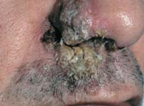
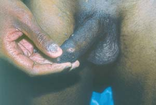
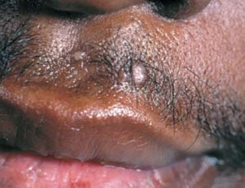
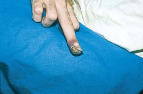 24.1 Herpes simplex. Shown here is HIV-associated chronic ulcerated herpes simplex that is resistant to acyclovir. This patient is receiving intravenous foscarnet. |
Basics
In the immunocompromised host, the clinical manifestations and course of herpes simplex virus (HSV) infection differ in patients with defective cell-mediated immunity, as seen in HIV infection. (See Chapter 6, “Superficial Viral Infections,” and Chapter 19, “Sexually Transmitted Diseases,” for a full discussion of HSV infections in immunocompetent hosts.)
Recurrent lesions may affect mucous membranes and possibly become chronic, centrifugally expanding ulcerations. These ulcerations last 1 month or more in an HIV-positive patient and are an AIDS-defining diagnosis.
Lesions may become resistant to acyclovir, or they may develop into chronic keratotic papules. Because acyclovir resistance is associated with prior treatment of suboptimal doses, it is important not to undertreat HIV-positive patients who also have HSV infections.
Description of Lesions
Initially, there are the typical grouped vesicles on an erythematous base, which evolve into pustules, erosions, and crusts.
Ultimately, the following lesions may occur:
Distribution of Lesions
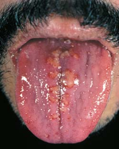
Intraoral areas, including the tongue, buccal mucosa, palate, and gingivae may be involved.
Chronic ulcerative lesions may appear in perianal areas. These lesions can extend into the intergluteal cleft (Fig. 24.4).
Keratotic lesions may occur in any location.
Clinical Manifestations
Lesions may be more severe and more extensive than in immunocompetent hosts.
Severe or chronic erosions, ulcerations, or keratotic lesions should alert the clinician to the presence of advanced immunosuppression.
Diagnosis
See Chapter 6, “Superficial Viral Infections,” and Chapter 19, “Sexually Transmitted Diseases,” for more detailed discussions.
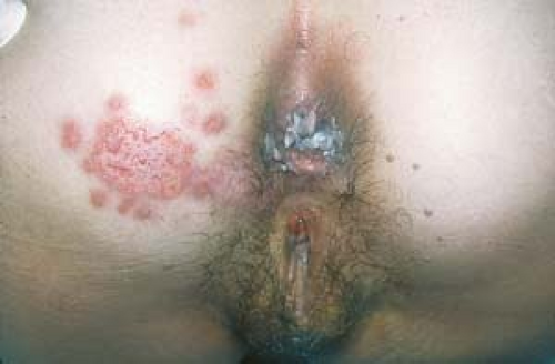 24.4 Herpes simplex. Chronic ulcerated lesions and scattered intact vesicles are present in a patient with AIDS. |
Herpes Zoster
Lesions of herpes zoster may involve only part of a dermatome and may be clinically indistinguishable from HSV lesions.
When in doubt, sufficient doses of antiviral medications are recommended for herpes zoster infection. Underdosing must be avoided.
Decubitus Ulcer
These lesions affect bony prominences in debilitated patients and do not extend to the intergluteal cleft.
Cutaneous Cytomegalovirus Infection
In cytomegalovirus infection, perianal ulcers develop as an extension of gastrointestinal involvement. Skin biopsy shows characteristic viral inclusion bodies.
In the keratotic type of cytomegalovirus infection, disseminated infection is associated with retinal findings, so an ophthalmologic examination is essential.
Disseminated Mycobacterium avium-intracellulare Complex
Patients may have oral ulcerations.
This infection is associated with severe systemic disease and fever in HIV-infected patients.
Disseminated Histoplasmosis
Patients may have oral and cutaneous ulcerations.
Disseminated histoplasmosis is associated with systemic disease.
If the patient has malabsorption or if lesions do not respond to other treatment, acyclovir (5 to 10 mg/kg every 8 hours) is infused over 1 hour. The dosage interval should be increased in patients with renal failure.
Failure to respond to intravenous acyclovir indicates acyclovir resistance.
Acyclovir resistance can be prevented by avoiding undertreatment and intermittent treatment.
Foscarnet (40 mg/kg intravenously every 8 hours) is used in acyclovir-resistant patients.
Strains that recur after treatment with foscarnet are usually acyclovir sensitive.
Long-term suppressive therapy with acyclovir has been associated with acyclovir resistance.
Treatment should continue until clinical lesions resolve completely.
Clinicians should be careful not to underdose with antiviral agents.
HIV-Associated Herpes Zoster
Basics
Herpes zoster is most common in elderly patients and in immunocompromised persons, although it may occur in anyone who has a history of chickenpox.
See Chapter 6, “Superficial Viral Infections,” for a more detailed discussion.
Description of Lesions
Grouped vesicles or bullae on an erythematous base affect all or part of a dermatome.
Lesions evolve into pustules and crusts and may erode. Chronic ulcerations and crusted or verrucous lesions may occur.
Severe scarring may result (Fig. 24.5).
Distribution of Lesions
Any dermatome can be affected.
Disseminated herpes zoster virus may occur.
Occasional dissemination may lead to 25 or more lesions outside of the primary and two contiguous dermatomes (Fig. 24.6).
The disease usually begins with typical dermatomal herpes zoster virus that becomes widespread and chronic.
The eruption may be indistinguishable from varicella.
Clinical Manifestations
Prodromal symptoms of pain and itching may be severe enough to lead to a suspicion of serious illness. For example, the prodromal pain of thoracic zoster has led to critical care unit admission to rule out myocardial infarction.
Regional adenopathy may occur.
Varicella pneumonia may develop.
Cutaneous lesions may become chronic in patients with AIDS.
Intravenous acyclovir (10 mg/kg every 8 hours) is given for 10 to 14 days.
Dosage intervals are increased in patients with renal failure.
If lesions improve but persist beyond 10 to 14 days, treatment is continued until all lesions resolve.
If lesions fail to resolve, the virus may be resistant to acyclovir.
Acyclovir-resistant varicella-zoster virus infection responds to foscarnet (40 mg/kg every 8 hours until lesions resolve).
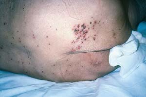 24.6 Disseminated herpes zoster. Note the initial dermatomal involvement on the buttock. (Courtesy of Herbert A. Hochman, M.D.) |
Undertreatment may lead to viral resistance.
Patients with herpes zoster can transmit the virus as chickenpox to nonimmune persons.
HIV-Associated Molluscum Contagiosum
Basics
Molluscum contagiosum is caused by a poxvirus; the condition is most commonly seen in immunocompetent children and less commonly in healthy adults.
Multiple and extensive facial lesions, as well as lesions with atypical morphology, should alert the practitioner to the possibility of HIV infection.
See Chapter 6, “Superficial Viral Infections,” for further discussion.
Description of Lesions
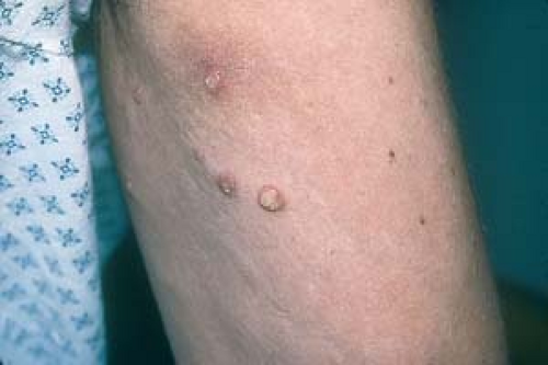
Papules may be dome-shaped or, more commonly, are atypical in appearance.
Size may be up to, or greater than, 1 cm (giant molluscum contagiosum).
Lesions may lack central umbilication or may have several umbilications.
Lesions on hairy areas tend to penetrate hair follicles.
Lesions may be extensive (hundreds to thousands in number) in patients with advanced AIDS.
Patients receiving HAART tend to have rare molluscum, with the more typical morphology seen in immunocompetent hosts.
The appearance of new lesions may follow a downward fluctuation in immunity caused by a concurrent infection, such as influenza.
Distribution of Lesions
Clinical Manifestations
There is occasional tenderness or inflammation.
Lesions are often a great cosmetic concern to patients.
Disseminated Cryptococcosis
Cutaneous lesions may be clinically identical to those of molluscum contagiosum (Fig. 24.9).
Affected patients are usually systemically ill, although cutaneous involvement may be the first sign of illness.
Crush preparation with India ink shows encapsulated yeast.
When in doubt, biopsy can be used to identify the lesions.
Patients with cutaneous dissemination have neurologic involvement, and a faster diagnosis can be made by cerebrospinal fluid examination.
Treatment is individualized for each patient. No specific treatment is universally more effective than any other.
Topical tretinoin is a useful adjunctive treatment in cases of molluscum contagiosum of the beard.
Surgical treatment is with curettage or liquid nitrogen cryosurgery.
Trichloroacetic acid 25% to 75% may be applied to individual lesions.
Podofilox (Condylox) 5% may be applied to lesions twice per day, 3 days per week.
Imiquimod 5% (Aldara) cream may be effective and should be used daily if possible.
Treatment is long term and is unlikely to eradicate all lesions unless the patient’s immunity improves. Lesions may remit spontaneously after the patient is started on HAART, and knowledge of this fact will sometimes persuade a reluctant patient to begin treatment for HIV infection.

