Fig. 42.1
Cutaneous leiomyosarcoma. A slow-growing erythematous to brownish nodule of 5 cm in diameter on the leg
The familial occurrence of cutaneous leiomyosarcoma with renal cancer has been described in the context of hereditary cutaneous leiomyomatosis and renal cell cancer (HLRCC). This rare genetic syndrome is caused by heterozygous mutations in the fumarate hydratase (FH) gene.
Pathology
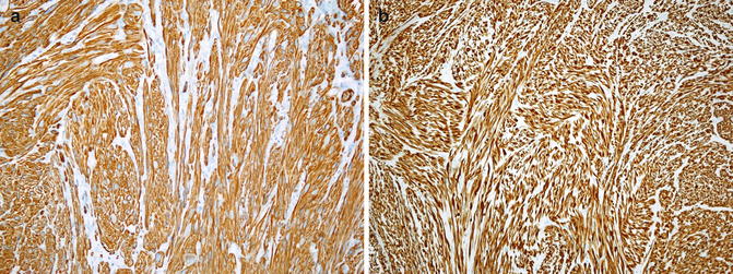
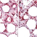
Superficial (dermal) leiomyosarcoma is confined to the dermis or showed only very superficial, focal subcutaneous extension. The neoplasm is characterized by a poorly circumscribed proliferation of interwoven fascicles of spindle-shaped atypical myomatous cells with eosinophilic cytoplasm and cigar-shaped nuclei that merge with a collagenous stroma (Figs. 42.2 and 42.3). Mitotic figures (1/2 per 10 high-power fields), high cellularity, and bizarre myomatous cells are the generally accepted criteria for malignancy (Fig. 42.4). Two growth patterns are described: a nodular pattern characterized by high cellularity, marked nuclear atypia, and numerous mitoses and a diffuse pattern that is less cellular and well differentiated and shows inconspicuous mitoses. Focal areas of hemorrhage with necrosis and inflammatory component are present. Unusual histological variants include epithelioid, granular cell, inflammatory, myxoid, and desmoplastic leiomyosarcoma. α-smooth muscle actin (α-SMA), muscle actin specific (HHF35), and calponin and caldesmon staining are positive in a vast majority of cases (Fig. 42.5a, b). However, none of these markers is absolutely specific for smooth muscle, and positivity for at least two of these markers is more supportive of leiomyosarcoma. Desmin is positive in 50–70 % of cases. MIB-1 labeling index can be useful as adjunctive aids for recognizing malignancy. Focal positivity for cytokeratins, EMA, CD34, and S-100 protein may be encountered. Although a loss of PTEN acting as a tumor suppressor gene has been reported in a case series, its significance in tumor genesis and its diagnostic utility remain to be determined.
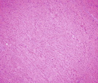
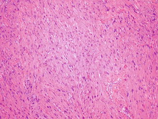
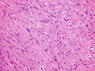
Fig. 42.2
A poorly circumscribed proliferation of interwoven fascicles of spindle-shaped atypical myomatous cells that merge with a collagenous stroma
Fig. 42.3
The cells show an eosinophilic cytoplasm and cigar-shaped nuclei
Fig. 42.4
Mitotic figures, high cellularity, and bizarre myomatous cells