Cutaneous Hematolymphoid Proliferations


Cutaneous Hematolymphoid Proliferations


Julia Scarisbrick

Louis P. Dehner

Alejandro A. Gru


INTRODUCTION

Primary cutaneous lymphomas are rare cutaneous neoplasms with varied clinical presentation and course. Initial examination must differentiate between primary cutaneous lymphoproliferative disorders (LPD) and secondary involvement of the skin by systemic lymphoma. In children, systemic lymphomas and leukemias are more common than primary cutaneous LPDs. Moreover, full staging investigations such as a bone marrow (BM) biopsy and imaging ensure accurate diagnosis of a primary skin lymphoma. The clinical course of primary cutaneous lymphomas differs from that of its systemic counterparts and requires a team-based approach involving hematologists, dermatologists, and other specialists. Morphologic interpretation of tumors involves the use of cytologic touch preparations, flow cytometry, fluorescence in situ hybridization (FISH), and conventional cytogenetics. The choice of molecular tools is of paramount importance as are the treatments that vary from an expectant/watchful waiting approach to aggressive chemotherapy regimens and possible allogeneic BM transplantation, depending on the histologic subtype of skin lymphoma.

Four major histologic subtypes account for most non-Hodgkin lymphomas (NHL) in the pediatric setting: Burkitt lymphoma (39%), diffuse large B-cell lymphoma (DLBCL; 16%), lymphoblastic lymphoma (28%), and anaplastic large cell lymphoma (ALCL; 10%).1 Approximately 7% of pediatric NHL includes more unusual histologic subtypes, including cutaneous lymphomas. The epidemiologic aspects of cutaneous hematopoietic tumors in children are poorly studied, as most of them are derived from small case series that are highly biased by private consultation material from single institutions.2 In the series by Fink-Puches et al3(n = 69), ˜35% of pediatric cutaneous lymphomas were accounted for by mycosis fungoides (MF), another ˜35% by CD30+ lymphoproliferative disorders (ALCL and lymphomatoid papulosis [LyP]), and 20% included marginal zone lymphomas (MZLs) and B-lymphoblastic lymphoma. The pediatric series by Boccara et al4 in France (n = 51) showed that nearly half of these tumors (47%) were composed by LyP, 27% by lymphoblastic lymphomas/ leukemias and leukemic infiltrates by acute myeloid leukemia (AML), and approximately 10% included cases of Epstein-Barr virus (EBV)+ lymphoproliferative neoplasms and MF.

Although most cutaneous lymphomas—with MF the most common—are similar in children and adults, a disproportionate number of lesions in children have a particular clinical and immunophenotypic profile. In children, most of the hypopigmented lesions have a CD8+ phenotype and exhibit a very indolent behavior. Similarly, lymphoblastic and myeloid leukemic infiltrates are overrepresented in children, as the diseases occur with relatively greater frequency. Histiocytoses with cutaneous presentation are more frequent in children than in adults.


Mature T-Cell and NK-Cell Lymphomas


PRIMARY CUTANEOUS ALCL, ANAPLASTIC LYMPHOMA KINASE (ALK)-NEGATIVE


Definition and Epidemiology

Primary cutaneous anaplastic large cell lymphoma (PC-ALCL) is composed of large and pleomorphic cells (indistinguishable from ALK+ ALCL) with diffuse expression of CD30 in the tumor cells (greater than 75%).5,6 The diagnosis is limited to those cases without a history of LyP or MF.7,8 Unlike ALK+ ALCL, PC-ALCL is very rare in children compared with adults9,10,11,12,13,14 and a diagnosis of LyP (type A or C) should first be considered. In some cases of PC-ALCL in childhood, an association with HIV infection has been documented.9


Clinical Presentation

Rapidly growing, asymptomatic, solitary or multiple skin nodules/tumors with a tendency to ulcerate (Figure 27-1) are the characteristic clinical presentation.12


Histologic Findings

Microscopically, ALCL has diffuse, cohesive sheets of large pleomorphic tumor cells, strongly CD30+, similar to ALK+ ALCL (Figures 27-2 and 27-3). Even among the conventional variants, a rich inflammatory infiltrate is present, which can lead to a misdiagnosis of an inflammatory process in the skin.15 The neutrophilic variant (pyogenic) of PC-ALCL contains a neutrophilic-rich infiltrate (Figure 27-4). Although the conventional variant does not have a predilection for immunocompromised individuals, the pyogenic variant is seen in those with HIV,16 transplant recipients,17 and hematologic malignancies, including young individuals.10,18


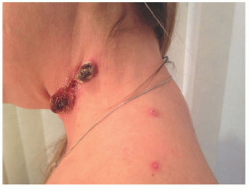
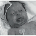
FIGURE 27-1. Cutaneous anaplastic large cell lymphoma of the neck in a girl with coexisting lymphomatoid papulosis. Large ulcerated necrotic tumors can be seen on the neck.

The tumors are frequently CD4+ and show expression of cytotoxic markers (TIA-1, perforin, granzyme B) (Figure 27-3). There is variable loss of T-cell antigens and, as opposed to systemic ALK+ ALCL, PC-ALCL is usually negative for epithelial membrane antigen (EMA).19 CD99 can also be positive.9 The pyogenic variant can have a higher rate of CD8 and EMA (57%) expression, compared to the conventional form.10,20 Tcell clonality has been proved in the vast majority of cases by conventional T-cell receptor (TCR) studies. ALK-ALCL have shown convergent mutations and kinase fusions that lead to constitutive activation of the JAK/STAT3 pathway, and a subset with rearrangements at the locus containing DUSP22 and IRF4 in chromosome 6p25 tends to be relatively monomorphic, usually lack cytotoxic granules, and have been reported to have a superior prognosis, whereas a small subset with TP63 rearrangements are very aggressive.21,22,23


Differential Diagnosis

The clinical differential diagnosis for the pyogenic variant of PC-ALCL includes pyoderma gangrenosum, pyoderma faciale (rosacea fulminans), Sweet syndrome, leishmaniasis, deep fungal infection, or pyogenic granuloma. Bacterial cellulitis has also been described as another differential diagnostic consideration. Among neoplastic conditions, LyP is common in children (see discussion below), and tumor stage MF, which typically presents in the fifth to sixth decade, is exceedingly rare in kids; the latter two disorders are best distinguished on a clinical background. Those with transformed MF have a long-standing history of the disease and present with multiple tumors, whereas LyP has a history of self-remitting papules. ALK staining and CD30 are useful to rule out the possibility of cutaneous involvement by systemic ALK+ ALCL.10


MYCOSIS FUNGOIDES


Definition and Epidemiology

MF is the most common form of cutaneous T-cell lymphoma in both adults24,25 and children.4,26,27,28,29 MF in children has a characteristic clinical appearance of hypopigmented


patches (Figure 27-5) and plaques that generally do not progress to the tumoral phase, and has a CD8+ phenotype.30 It has been reported in children as young as 3 years.3,27,31,32,33,34 The male-to-female ratio is nearly equal in the pediatric setting.27,31,34,35 Fink-Puches et al showed that MF represented almost 35% of all cutaneous lymphomas in individuals younger than 20 years.3


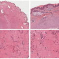
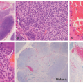
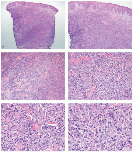
FIGURE 27-2. Cutaneous anaplastic large cell lymphoma—histopathologic findings. Diffuse dermal infiltrate sparing the surface epidermis (A and B). The malignant infiltrate dissects through the collagen bundles (C). There is a rich admixed neutrophilic inflammatory infiltrate (D). The infiltrate is composed of malignant appearing large cells with many “hallmark” cells. Eosinophils are also seen (E and F).


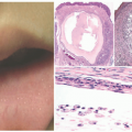
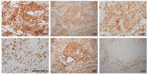
FIGURE 27-3. Cutaneous anaplastic large cell lymphoma—immunohistochemistry. The malignant cells are strong and diffusely positive for CD30 (>75%), CD4, CD3, and have patchy positivity for granzyme B. They have retained expression of CD5, whereas there is a marked loss of CD7.


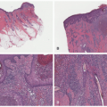
FIGURE 27-4. Cutaneous anaplastic large cell lymphoma, pyogenic variant. The very dense infiltrate is obscured by a rich acute inflammatory infiltrate with neutrophilic and numerous eosinophils (A-C). The malignant cells are unmasked by a CD30 stain.


FIGURE 27-5. Mycosis fungoides in an adolescent. Characteristic hypopigmented patches can be seen on the lower back.


Etiology

The discovery of specific molecular rearrangements36,37,38,39,40 complements advances in the mutational landscape of MF and Sézary syndrome (SS). Choi et al36 found frequent deletions in chromatin modifying genes (ARID1A [62.5%], CTCF [12.5%], and DNMT3A [42.5%]). RB1 was deleted in 25% of samples. CARD11 and JAK2 amplification were seen in 22.5% and 12.5% of cases, respectively. MYC amplification was noted in 42.5% of samples. da Silva Almeida et al37 identified a median of 21 copy number alterations per sample (range 0-56) in SS, with characteristic recurrent gains in chromosomes 7 (5/25, 20%), 8q (13/25, 52%), and 17q (2/25, 8%), as well as recurrent deletions involving tumor-suppressor genes in 17p13.1 (TP53; 13/25, 52%), 13q14.2 (RB1; 4/25, 16%), 10q23.3 (PTEN, 5/25; 20%), and 12p13.1 (CDKN1B; 5/25, 20%). Kiel et al38 showed numerous (n = 42) fusion genes including TPR-MET, MYBL1-TOX, DNAJC15-ZMYM2, and EZH2-FOXP1, which, albeit not recurrent, could contribute to the pathogenesis of SS. Ungewickell et al39 found structural variation events (excluding copy number gains) in pathways related to T-cell survival and proliferation in 11% of patients with MF or SS. The structural variants were largely mutually exclusive with the TNFR2 alterations. The structural variants included NFKB2 gene truncations in 5% (4/73) of cases with the deletion of a region whose loss generates a truncated p100 protein with predicted proteasome-independent NF-κB2 nuclear localization, as well as a deletion involving TRAF3 that increases noncanonical NF-κB signaling. A recurrent CTLA4-CD28 fusion was also discovered.


Clinical Presentation

Clinically, pediatric and adult MF may be difficult to diagnose, partly because they can potentially mimic numerous benign inflammatory dermatoses (Figure 27-6). In fact, a diagnosis is commonly delayed and may take several years, and numerous biopsies are often required to eventually establish or consider the diagnosis before clonality studies are performed. Ackerman raised concerns about the diagnosis of MF in the pediatric setting in the past: in their review of 106 cases, it was claimed that only 23 cases had information “sufficient” for a diagnosis of hypopigmented MF.41 Indeed, in 83 of those cases, there was no significant clinicopathologic correlation to confidently establish the diagnosis.

MF also enters into the differential diagnosis of psoriasis, tinea corporis, pityriasis lichenoides (PL), lichen aureus, atopic dermatitis, and the various hypopigmented dermatoses. Vitiligo and pityriasis alba are the two most frequent misdiagnoses in children.42,43 In a recent case series, all children (100%, n = 69) presented at the patch stage; almost all cases had hypopigmented lesions and most of these occurred in African American children.26 Compared with children, adults exhibit sharply demarcated, atrophic lesions with a cigarette paper appearance (Figures 27-7 and 27-8). In an earlier study by Crowley et al, of the 58 patients with MF (younger than 35 years), 17% presented with the tumor stage and approximately 4% had generalized erythroderma.27 In another series of 46 cases, Heng et al reported that over 90% had hyperpigmented lesions.44 The most commonly affected areas included the buttocks, trunk,
and extremities (sun-protected areas). About 6% of patients have solitary lesions. Rare cases of MF can present following organ transplantation.45


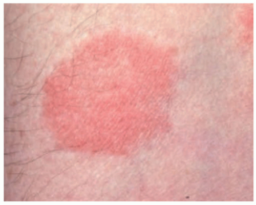
FIGURE 27-6. A patch of mycosis fungoides may be similar to other more common childhood dermatoses such as eczema or psoriasis.


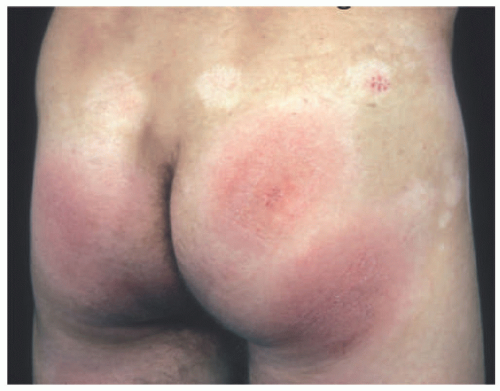
FIGURE 27-7. Distribution of patches of mycosis fungoides is more common in the “bathing suit” area.

Folliculotropic MF (FMF) presents with follicular papules occasionally with an erythematous base, and follicular plugging and/or alopecia; this is the second most common clinical variant and seemingly occurs more frequently in individuals younger than 40 years (Figure 27-9) (8% of all variants).46,47,48,49,50 In FMF, the lesions are usually located in the head and neck region, with the presentation of plaques and/or tumors and intense pruritus. FMF has been associated with a worse outcome in adults and children unlike the more indolent and common patch/plaque lesions without FMF.46 Less common clinical variants in the pediatric population include “granulomatous slack skin” or granulomatous MF,51,52,53,54,55 characterized by the development of areas of pendulous, lax skin in the major skin folds (especially axillae and groins).56 Localized pagetoid reticulosis (PR; Woringer-Kolopp disease) presents as a solitary, slowly growing erythematous and verrucous plaque on the extremities; rare cases have been reported in children.57,58,59 SS is a distinctive erythrodermic cutaneous T-cell lymphoma, characterized by pruritic erythroderma, generalized lymphadenopathy, and circulating malignant cells with cerebriform nuclei. It is a disease of adults, with only rare cases in the pediatric population.60,61,62 A rare clinical presentation of palmoplantar keratoderma has also been described in children.63


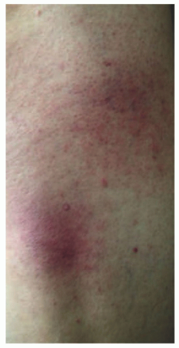
FIGURE 27-8. Patches of mycosis fungoides show poikilodermatous skin change with atrophy, telangiectasia, and dyspigmentation.


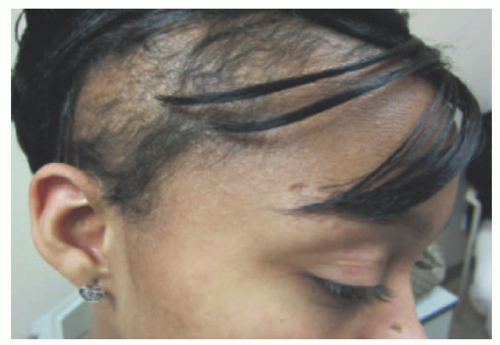
FIGURE 27-9. Mycosis fungoides, folliculotropic form. Patches of alopecia in an adolescent girl.


Histologic Findings

Morphologically, pediatric MF is histologically indistinguishable from that seen in adults (Figure 27-10, 27-11, 27-12 and 27-13). According to the series from Castano et al, the most frequent histologic findings include: lymphocytes in clusters, patchy lichenoid infiltrates, perivascular/periadnexal infiltrates, psoriasiform hyperplasia, papillary dermal fibroplasia, dermal melanophages, and Pautrier microabscesses. The infiltrate shows epidermotropism with tagging of cells along the dermal-epidermal junction.64 The atypical lymphocytes show nuclear hyperchromasia and irregular and sometimes cerebriform nuclei. Distinctive perinuclear halos are seen surrounding the intraepidermal lesional cells. Admixed histiocytes, plasma cells, and eosinophils can be present. As the lesions progress clinically, so does the extent of the infiltrate. Large cell transformation is defined as the presence of large cells (at least 4 times the size of a small lymphocyte) comprising >25% of the infiltrate or in


nodular aggregates. Only rarely has such a phenomenon been described in lesions in children.65,66 In FMF, there is infiltration of the hair follicle epithelium with or without epidermotropism (epidermotropism is more frequently seen). The infiltrate involves the infundibulum of the hair follicle and at times deeper portions of the follicle. Follicular mucinosis may be an accompanying feature that can be better demonstrated by colloidal iron or Alcian blue stains. Additionally, FMF is often accompanied by a syringotropic infiltrate.46 However, a Dutch study revealed that interfollicular epidermotropism is actually rare.50 FMF is not usually accompanied by intraepidermal Pautrier microabscesses.67 Dermal eosinophilia can be prominent, particularly during the progression of the disease, and might be a manifestation of an autoimmune response to the keratin of the hair shafts in the dermis. In granulomatous MF (GMF), dense nodular and diffuse granulomas are present in the dermis, with or without epidermotropism, and with destruction of elastic fibers and elastophagocytosis. In the PR variant of MF, prominent pagetoid epidermotropism is noted (Figure 27-14). Such cases show a very impressive clinical response to radiation treatment.


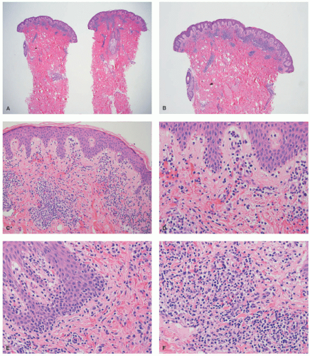
FIGURE 27-10. Pediatric hypopigmented mycosis fungoides. (A and B, low magnification—20× and 40×). There is a superficial dermal band-like infiltrate. (C and D, intermediate magnification, 100× and 200×). The infiltrate is associated with extravasation of red blood cells. There is tagging of lymphoid cells at the dermal-epidermal junction. (E and F, high magnification, 400× each). The infiltrate is composed of small- to medium-sized lymphocytes, with hyperchromasia, and irregular nuclear borders. The epidermotropic cells do not reveal definitive intraepidermal collections of lymphocytes in the form of Pautrier microabscesses. Reprinted with permission from Gru AA, Schaffer A. Hematopathology of the skin: A clinical and pathologic approach. 1st ed. Philadelphia, PA: Wolters Kluwer; 2017.


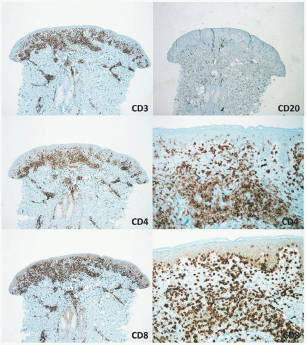
FIGURE 27-11. Pediatric hypopigmented mycosis fungoides—immunohistochemistry. CD3 is positive in the majority of the dermal and epidermal lymphocytes. CD20 is predominantly negative. CD4 shows positive staining in many of the T cells and the dermal lymphocytes. However, the vast majority of T cells in the epidermis are negative for CD4 and positive for CD8, which reveals extensive epidermotropism and tagging of cells along the dermal-epidermal junction. Reprinted with permission from Gru AA, Schaffer A. Hematopathology of the skin: A clinical and pathologic approach. 1st ed. Philadelphia, PA: Wolters Kluwer; 2017.


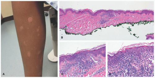
FIGURE 27-12. Pediatric mycosis fungoides, hypopigmented variant. Ill-defined hypopigmented patches are present in the leg. Histopathologically, there is an atypical infiltrate with epidermotropism. Tagging of lymphocytes along the dermal-epidermal junction is seen. The lymphocytes are small, with hyperchromasia, irregular nuclear borders, and perinuclear halos. Obtained with permission Gru A, Dehner LP. Cutaneous hematolymphoid and histiocytic proliferations in children. Pediatr Dev Pathol. 2018;21(2):150-207.


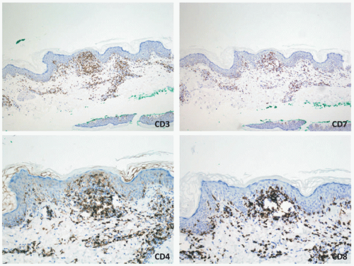
FIGURE 27-13. Pediatric mycosis fungoides, hypopigmented variant—immunohistochemistry. The neoplastic cells are positive for CD3 and CD8. The CD4:CD8 ratio is inverted. There is aberrant loss of CD7. Obtained with permission Gru A, Dehner LP. Cutaneous hematolymphoid and histiocytic proliferations in children. Pediatr Dev Pathol. 2018;21(2):150-207.

The immunophenotype of pediatric MF is different from most of the adult forms (Figures 27-11 and 27-13): hypopigmented MF is a CD8+ cytotoxic T-cell lymphoma (whereas most cases in adults are CD4+).3,26,27,28,29,31,32,41,42,44,46,68,69,70,71,72,73,74,75 Also, CD30 expression is usually negative or reacts with only a few scattered cells (80% of cases are entirely nonreactive). Decreased expression of both CD4 and CD8 can also be seen in some cases of hypopigmented MF.76,77 Similar to adult MF, there is usually preserved expression of CD2 and CD5 with loss of CD7. A single rare case with co-expression of CD56 has been reported with an associated indolent clinical course.78 In FMF the infiltrate usually shows a predominance of CD4+ cells like the adult cases.46 GMF is also a disease of CD4+ T cells. The genetics of MF and SS is less well understood in the context of the pediatric cases when compared with adult MF. T-cell gene rearrangement studies performed in pediatric MF revealed TCR clonality in approximately 64% of cases and a polyclonal result in 16% of cases. These numbers appear to be lower when compared to the adult experience of 80% clonality, which may reflect a relatively lower number of neoplastic cells in skin biopsies from children. However, sensitivity for TCR is lower in early lesions of MF (up to 60%).79 Hodak et al revealed monoclonality in only 43% of cases.46


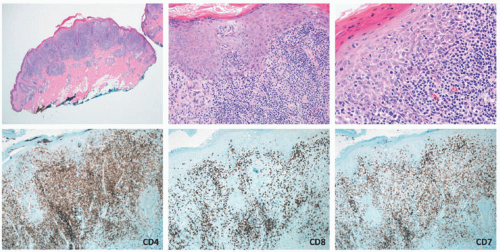
FIGURE 27-14. Pediatric mycosis fungoides, pagetoid reticulosis variant. In this case, the infiltrate shows very extensive pagetoid epidermotropism. Lymphoma cells are CD4 positive, CD8 negative, and have loss of CD7. Obtained with permission Gru A, Dehner LP. Cutaneous hematolymphoid and histiocytic proliferations in children. Pediatr Dev Pathol. 2018;21(2):150-207.


Differential Diagnosis

The differential diagnosis of MF includes a number of inflammatory dermatoses: Pityriasis alba usually presents with hypopigmented macules and patches on the face but may also affect the trunk and limbs. Histologically, there are very sparse lymphoid infiltrates, with very slight spongiosis and without the dermal fibrosis seen in MF; vitiligo consists of depigmented macules and patches in localized, segmental, or widespread distribution. The lesions are usually on the face and distal extremities. Histologically, the findings are minimal, and if present, there is a subtle perivascular infiltrate of lymphocytes with minimal epidermal involvement. As lesions evolve, there is a decrease in the number of
melanocytes at the dermal-epidermal junction, which can be proved with the use of a MART-1 immunostain. Acral pseudolymphomatous angiokeratoma of children (APACHE) can also mimic PR. A dense lymphoid infiltrate beneath the epidermis, and thick wall vessels are seen.80 PL chronica and varioliformis acuta (PLC/PLEVA) can also share histomorphologic features. A lymphocytic vasculitis and interface changes are present in both conditions. As opposed to MF, loss of T-cell antigens (CD7) is not typical. Molecular studies (TCR rearrangement) should not be used to distinguish between both conditions, particularly if the BIOMED primers are being used, because PLC and PLEVA can have positive clonality studies in up to 25% of cases. Next generation sequencing accurately distinguishes between inflammatory dermatoses with clonal populations of cells and cutaneous T-cell lymphomas.81 However, this technique is currently expensive and unlikely to replace TCR gene analysis in the clinical setting. Spongiotic dermatoses (eczema, contact dermatitis, etc) can also enter the differential diagnosis. The latter typically lack significant cytologic atypia of the lymphoid population or aberrant antigenic loss. If the infiltrate is positive for CD30, other CD30+ lymphoproliferative diseases (LPDs) can enter the differential diagnosis (LyP and PC-ALCL). In children, Langerhans cell histiocytosis is also accompanied by extensive pagetoid epidermotropism of the histiocytic cells. As opposed to MF, langerhans cell histiocytosis (LCH) is positive with histiocytic markers (CD68, S100, CD1a, and Langerin), whereas it is negative for T-cell antigens. A word of caution should be made with LCH, as many cases can have clonal rearrangements of the TCR.


LYMPHOMATOID PAPULOSIS


Definition and Epidemiology

Lymphomatoid papulosis (LyP) is a CD30+ T-cell lymphoproliferative disorder characterized by recurrent crops of papulonecrotic lesions that usually prevail for 3 and 8 weeks and then resolve spontaneously.82 The lesions are clinically “benign” appearing, but the histologic features suggest otherwise. However, the lesions may frequently heal with atrophic scarring, which may be disfiguring. LyP is the third most common type of cutaneous lymphoproliferative disorder in children.3,83 Boys are reported to have an earlier onset of LyP compared with girls. It is estimated that between 5% and 20% of those with LyP can develop a subsequent lymphoma, usually MF, Hodgkin lymphoma, or ALCL; this proportion appears to be smaller in the pediatric setting (approximately 9%).83,84,85,86,87,88 The frequency of LyP ranges from 16% to 47% of all skin lymphomas in children, across different case series.2,3,4


Etiology

Karai et al were the first to report a group of patients with LyP and associated rearrangements of the IRF4-DUSP22 locus on 6p25.3.89 The patients showed unusual histologic findings, including PR-like epidermal changes with a proliferating dermal tumor. A consistent histologic finding also included a typical periadnexal infiltrate and hallmarklike cells.


Clinical Presentation

Clinically, LyP presents with erythematous papules and nodules that become hemorrhagic and necrotic after a few days with resolution as a depigmented scar.4,7,71,82,83,84,85,86,87,90,91,92,93 Lesions at different stages of evolution are characteristically present (Figure 27-15). In children, there is a clinical resemblance to PL, the most important disease in the differential diagnosis. The sites of predilection are the trunk and extremities, but other locations have been reported.93 Regional LyP with crops of lesions located in a single anatomic region is seemingly more common in children.92,93 Nearly 50% of cases in children have more than 50 lesions at one point during their clinical course.

LyP is a clinical diagnosis of self-resolving papulonecrotic lesions. Although most are CD30 positive, the histology may be varied and indistinguishable from transformed CD30-positive MF, popular MF, or PC-ALCL in some cases.


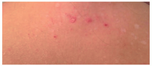
FIGURE 27-15. Lymphomatoid papulosis (LyP). Lesions of LyP often appear in crops; they mature from red papules and may ulcerate and tend to heal with atrophic hypopigmented scars.


Histologic Findings

Several histologic variants have been described in LyP: types A, B, C, D, E, and F (Table 27-1). None of the histologic subtypes have any prognostic significance. The importance in differentiating the various subtypes is related to the differential diagnosis that each subtype entails. Many of these lesions have substantial histopathologic overlap: Type A LyP has a wedge-shaped dermal infiltrate composed of medium to large and pleomorphic cells scattered throughout the infiltrate or are arranged in clusters (Figure 27-16). The malignant cells can show features of Hodgkin-like cells, and there is a rich background of inflammatory cells. Ulceration, edema of the dermis, and focal vasculitic changes may be present. Type B LyP is
characterized by an epidermotropic infiltrate reminiscent of MF, whereas Type C LyP has features histologically identical to PC-ALCL. Clearly the clinical distinction is more important in a sense than the histologic findings. Cerroni et al showed that a majority of the children represented types A and C, with no examples of type B. In another pediatric series of 250 cases LyP in children, most cases belonged to type A.88 Type D LyP mimics aggressive epidermotropic CD8+ T-cell lymphoma histologically94; this variant expresses other cytotoxic markers such as granzyme and TIA-1. In the latter study, 4 of the 9 patients were less than 25 years of age. Type E LyP is characterized by an angioinvasive and angiodestructive CD8+ cytotoxic T-cell infiltrate with CD30 co-expression; 2 of the 16 original patients were children.95 Another study of 14 children with LyP revealed that 10 cases had a CD8+ cytotoxic phenotype; most cases were considered either type A or type C. Pierard et al introduced the term follicular LyP (type F) to describe a subtype with a predominant perifollicular pattern of infiltration.96,97 A few cases of LyP (type F) have been reported in children.98,99


TABLE 27-1. Lymphomatoid Papulosis, Subtypes


LyP Type


Histologic Findings


Differential Diagnosis


Criteria


Type A


  • Wedge-shaped dermal infiltrate


  • Scattered or in small clusters arranged large CD30+ lymphocytes with nuclear pleomorphism and mitotes


  • Background infiltrate of histiocytes, eosinophils, neutrophils.


  • MOST COMMON IN CHILDREN


  • Hodgkin lymphoma (primary or secondary cutaneous)


  • MF (transformation)


  • Staging examination (in nodal HL)


  • Patches and plaques in MF vs selfregressing papulonodular lesions in LyP


Type B


  • Epidermotropic infiltrate of small- to medium-sized lymphocytes with atypical chromatin-dense nuclei


  • Variable expression of CD30 (0%-77%)


  • MF (patch/plaque stage)


  • Cutaneous γ/β lymphoma (epidermotropic)


  • Patches and plaques in MF vs selfregressing papulonodular lesions in LyP


  • Multiple plaques and nodules with ulceration.


  • IHC: Expression of TCRγ


Type C


  • Nodular cohesive infiltrate of large CD30+ pleomorphic or anaplastic lymphocytes with abundant cytoplasm and mitotic activity.


  • Admixture of only few eosinophils and neutrophils.


  • Anaplastic large cell lymphoma (primary cutaneous or systemic form)


  • MF (transformation)


  • Peripheral T-cell lymphoma, NOS (primary cutaneous or nodal)


  • Adult T-cell lymphoma/leukemia


  • Clinical presentation with solitary or grouped nodules in PC-ALCL. Staging examinations in SALCL. Patches and plaques preceding tumors in MF. Lack of CD30 or expression by only a minority of tumor cells (usually <30%), Staging examinations


  • Integration of HTLV-1/2 in tumor cell genome.


Type D


  • Epidermotropism of atypical small- to medium-sized pleomorphic lymphocytes


  • Deep dermal or subcutaneous perivascular infiltrates may be present.


  • Expression of CD8 (100%) and CD30 (90%)


  • 4/9 CASES IN CHILDREN


  • Pagetoid reticulosis


  • Primary cutaneous aggressive epidermotropic CD8+ cytotoxic T-cell lymphoma


  • Cutaneous γ/δ lymphoma


  • Unilesional erythematous scaling lesion in PR (eczematous or psoriasiform clinical aspect)


  • Multiple rapidly evolving plaques and nodules with erosions and necrosis.


  • No expression of CD30!


  • Multiple plaques with erosions IHC: expression of TCRγ


Type E


  • Angioinvasive, ie, angiocentric and angiodestructive infiltrates of mostly medium-sized pleomorphic CD30+ lymphocytes.


  • Vascular occlusion by atypical lymphocytes and/or thrombi, hemorrhage, extensive necrosis and ulceration.


  • Admixture of eosinophils.


  • Expression of CD8+ (70%) and CD30 (100%)


  • 2/16 CASES IN CHILDREN


  • Extranodal NK/T-cell lymphoma, nasal type


  • Cutaneous gamma/delta lymphoma


  • Anaplastic large cell lymphoma (primary cutaneous or systemic form) with angiocentric and angiodestructive growth)


  • Association with EBV, mostly secondary cutaneous involvement (staging)


  • IHC: Expression of TCR gamma


  • Clinical presentation with solitary or grouped nodules in PC-ALCL. Staging examinations in SALCL.


Abbreviations: HL, Hodgkin lymphoma; IHC, immunohistochemistry; LyP, lymphomatoid papulosis; MF, mycosis fungoides; NOS, nodal or systemic malignant; PC-ALCL, primary cutaneous anaplastic large cell lymphoma; SALCL, systemic anaplastic large cell lymphoma.


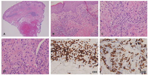
FIGURE 27-16. Lymphomatoid papulosis (LyP), type A. In LyP, there is a wedge-shaped infiltrate with only partial epidermal involvement (A and B). Malignant large and pleomorphic cells are present with a rich acute inflammatory (C and D) background. The lesional cells are positive for CD30 and have significant loss of CD3.

The immunophenotype of types A and C is very similar with a uniform population of CD30+ cells, with frequent CD3 and CD4 co-expression. Typically, CD8 and CD56 are negative. CD 15 is usually negative but cytotoxic molecules (TIA-1 and granzyme) are positive.100,101,102 Types D and E have a CD8+ phenotype. MUM1 is frequently expressed in all cases of LyP (but also in MF and PC-ALCL). Some cases of LyP can also have a DUSP22 translocation.89


Differential Diagnosis

Arthropod bite reactions should be particularly considered in the differential diagnosis. Arthropod bites tend to be more itchy and do not heal with scarring. Those can also have a wedge-shaped pattern histologically of inflammation as seen in type A LyP. As opposed to LyP cases, arthropod bites never have the number of CD30+ cells, or the aberrant loss of T-cell antigens. Pseudolymphomas should also be distinguished from LyP. Although the density of the infiltrate can be similar, the number of CD30+ cells is lower in pseudolymphomas, and they also lack T-cell antigenic loss. PC-ALCL can also be seen in children, and the main distinction appears to be clinical rather than pathologic. PC-ALCL tends to be more solitary, persists for a longer period, and less frequently shows spontaneous resolution. At the molecular level, both can have the DUSP22 translocation.


ANAPLASTIC LARGE CELL LYMPHOMA, ALK-POSITIVE (ALK+ ALCL)


Definition and Epidemiology

ALK+ ALCL represents approximately 10% to 30% of childhood lymphomas and is more frequent in the first three decades of life. There is a slight male predominance, and the majority of patients (70%) present with stage III or IV disease and B symptoms. The skin is the most frequent extranodal site of involvement (present in 26% of cases). Other extranodal sites include bone, soft tissues, lung, and liver.103,104,105,106 ALK+ ALCL has a good prognosis with a 5-year survival rate of 70% to 80% in contrast to 49% and 32% 5-year survival rates for ALK-negative ALCL and peripheral T-cell lymphoma, not otherwise specified (PTCL, NOS), respectively.107,108


Etiology

ALK+ ALCL has rearrangements of the ALK gene on 2p23 with various partner genes, most typically the nucleophosmin (NPM) on 5q35.109,110 Some translocations can be cryptic by conventional cytogenetic methods. Other translocation partners include nonmuscle tropomyosin (TPM3, 1q25 and TPM4, 19p13.1); amino terminus of 5-aminoimidazole-4-carboxamide ribonucleotide formyltransferase/IMP cyclohydrolase gene (ATIC, 2q35); TRK-fused gene (TFG, 3q21); clathrin heavy polypeptide gene (CLTC, 17q23); moesin gene (MSN, Xq11-12); myosin heavy chain 9 gene (MYH9, 22q11.2); and ALK lymphoma oligomerization partner on chromosome 17 (ALO17, 17q25).105,111


Clinical Presentation

Most cases of ALK+ ALCL present with lymphadenopathy. The most common extranodal site is the skin (26%).112,113,114,115 Other affected sites include bone, lung, liver, and soft tissues.105 A leukemic presentation is rare, and more frequent in the small cell variant.116 The BM is affected in a small percentage (10%-30%) of cases.117

The cutaneous manifestation of ALCL, ALK+ can be precipitated by insect bites.118,119 Lesions resemble typical arthropod bite reactions without clinical resolution. Most patients with ALCL, ALK+, or ALK- have pink papulonodular lesions.120,121,122 The lesions are more frequently solitary and rarely multiple. Rare cases of isolated cutaneous ALCL, ALK+ have been reported.123 Some uncommon clinical appearances include generalized erythroderma,124 orbital lesions,125 and ichthyosis.126


Histologic Findings

ALK+ ALCL has a variety of morphologic presentations. All cases show a malignant population of large cells with eccentric horseshoe or kidney-shaped nuclei with an eosinophilic region near the nucleus, referred to as “hallmark” cells. In some variants, the hallmark cells can be small. Occasionally, the malignant cells can mimic Reed-Stenberg cells or their variants. There are five distinctive variants of ALK+ ALCL: common, small cell variant, Hodgkin-like pattern, lymphohistiocytic pattern, and combined forms.111,123,127,128,129 Any of these patterns may present in the skin (Figures 27-17 and 27-18). The immunophenotype of ALK+ ALCL includes CD30 and ALK expression; the pattern of CD30 expression is both Golgi and cytoplasmic. Most cases are positive for EMA, TIA-1, granzyme B, and perforin, and show loss of T-cell antigens such as CD3, CD5, and CD7. Some cases can be null for all T-cell markers. CD43, CD2, and CD4 are more frequently positive.

The cellular localization of the ALK expression correlates with the pattern of translocation: the NPM/ALK fusion leads to both nuclear and cytoplasmic ALK staining. The ALK staining patterns in less common translocation variants include diffuse cytoplasmic (eg, TPM3, ATIC, TFG, TPM4, MYH9, ALO17), granular cytoplasmic (CLTC) (Figure 27-18), or membranous (MSN). ALCL, ALK- shows strong and diffuse expression of CD30. The pattern can be membranous, Golgi, and/or cytoplasmic. The strong and diffuse character is often a helpful clue to distinguish ALCL, ALK- from PTCL, NOS. Lack of multiple pan T-cell antigens and the expression of cytotoxic markers are characteristic. EMA and clusterin can be positive, but less frequently than in ALCL, ALK+.108

Lamant et al suggested a possible relationship between insect bites and the development of ALK+ ALCL118; this series of five patients had recent arthropod bites and developed nodal disease in the area of the skin lesions. Two of the skin biopsies revealed the presence of ALK+ cells. A complete remission was obtained after chemotherapy in all but one patient, who developed progressive disease and died. Oschlies et al123 described a series of six children, within the context of 487 patients enrolled in the Anaplastic Large Cell Lymphoma-99 trial with disease limited to the skin; these patients had complete remissions on follow-up, and most of them were treated with surgical excision only. In all but one patient, the pattern of ALK staining was nuclear and cytoplasmic, and FISH was positive for the ALK-NPM translocation. Clinically, in 1 of 6 patients the lesions were solitary (maculopapules or nodules), and 1 patient had multiple lesions. Previous isolated case reports of cutaneous presentations of ALK+ ALCL have been described.113,130,131 Despite such reports, most cases of ALK+ ALCL have associated systemic disease and require full staging.


Differential Diagnosis

From the diagnostic perspective, a diagnosis of ALK+ ALCL implies the presence of a systemic lymphoma “until proven otherwise.” The rare isolated cutaneous forms are very infrequent. All patients should invariably undergo extensive

staging procedures. The presence of ALK+ distinguishes this process from PC-ALCL. More recently, molecular studies have proved that systemic ALK-ALCL with the presence of DUSP22 translocations has a clinical outcome similar to cases of ALK+ ALCL. Inflammatory myofibroblastic tumors in children can also be ALK+. As opposed to ALK+ ALCL, such tumors are positive only for histiocytic markers and lack the cellular pleomorphism that is typical of ALCL cases.


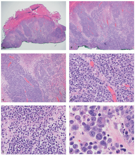
FIGURE 27-17. ALK+ Anaplastic large cell lymphoma with initial skin presentation. A dense malignant infiltrate in the dermis with surface ulceration, but sparing of the epidermis (A and B). The infiltrate shows angiotropism (C and D). It is composed of malignant large cells with frequent hallmark forms (E and F). Adapted and obtained with permission Gru AA, Voorhess PJ. A case of ALK+ anaplastic large cell lymphoma with aberrant myeloperoxidase expression and initial cutaneous presentation. Am J Dermatopathol. 2018;40(7):519-522.


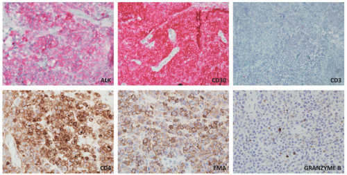
FIGURE 27-18. ALK+ Anaplastic large cell lymphoma with initial skin presentation. The large cells are positive for ALK (cytoplasmic granular staining), CD30, CD4, EMA, and weakly for granzyme B. They are negative for CD3. Adapted and obtained with permission Gru AA, Voorhess PJ. A case of ALK+ anaplastic large cell lymphoma with aberrant myeloperoxidase expression and initial cutaneous presentation. Am J Dermatopathol. 2018;40(7):519-522.


SUBCUTANEOUS PANNICULITIS-LIKE T-CELL LYMPHOMA


Definition and Epidemiology

Subcutaneous panniculitis-like T-cell lymphoma (SPTCL) is a mature T-cell lymphoma of the skin with TCR αβ expression, subcutaneous involvement, and sparing of the epidermis. It most commonly involves the extremities, occurs in different age groups (including children), and has an indolent clinical behavior.132 SPTCLs with expression of γδ TCR have distinct clinicopathologic features and are currently classified under γδ T-cell lymphomas.133 It is more common in women and has a predilection for younger individuals. The median age at presentation is 36 years (range 1-79). Approximately 50% of cases occurred in individuals 21-40 years old, and 19% of cases are 20-year-old or younger patients.134 Numerous cases have been reported in children.134,135,136,137,138,139 A subsequent study of cutaneous lymphomas in children noted that SPTCL was rare, with only 3.4% of patients in this age group.71


Etiology

The etiology of SPTCL remains unknown. Nearly 19% of patients with SPTCL have associated autoimmune disorders, including systemic lupus erythematosus, juvenile rheumatoid arthritis, Sjögren disease, type 1 diabetes mellitus, idiopathic thrombocytopenia, multiple sclerosis,
Raynaud disease, and Kikuchi disease.134 Yi et al140 showed a series of 11 cases of SPTCL initially diagnosed as autoimmune disorders; the authors divided the original diagnoses into three separate groups: (1) a group with a preceding diagnosis of erythema nodosum, pyoderma gangrenosum, lupus profundus; (2) vasculitis; (3) inflammatory myopathy-like lesions. The authors speculated that patients with inflammatory myositis and/or Behcet’s represented paraneoplastic manifestations of SPTCL. Some of these patients (2/11) had elevation of antinuclear antibodies (ANA) in the serum, anti-DS DNA antibody, and one patient had an elevated antineutrophil cytoplasmic antibody. A study from the Mayo Clinic on a series of 23 patients revealed preceding diagnoses of autoimmune disorders in approximately 57% of patients, including lupus panniculitis, erythema nodosum, venous stasis, Weber-Christian disease, cellulitis, and granulomatous panniculitis of unknown etiology. The association between SPTCL and lupus erythematosus profundus (LEP) has led to the hypothesis that perhaps the two entities represent two ends of the same spectrum.141,142 Some cases of lobular panniculitis with a CD8+ phenotype can occur in children in association with clonal populations of T cells in the setting of congenital immune deficiency syndromes. Rare cases in association with HIV143 and sarcoidosis have also been documented.144 The association between certain medications and development of SPTCL has been described in patients receiving anti-TNFα therapy (etanercept),145 and following rituximab and cyclophosphamide.146 More unusual presentations have been reported in patients with Down syndrome,147 cervical cancer,148 during pregnancy,149 and neurofibromatosis type 1.150


Clinical Presentation

The typical lesions consist of nodules/tumors or skin plaques, which can vary in diameter from 1 to 20 cm or more and are frequently multifocal (78%). Sometimes the nodules leave areas of lipoatrophy after resolution. Ulceration is rare (6%). The lesions present in the extremities (legs, arms) and, less frequently, on the trunk and face. Facial lesions can present with extraocular muscle palsy.151 Rare cases in the breast have been reported.152,153 The skin lesions often simulate other causes of panniculitis, such as EN154 and lupus panniculitis. Other rare clinical presentations might simulate dermatomyositis,155,156 morphea,157 cellulitis,158 facial edema,159 venous stasis-like ulceration,160 lipomembranous panniculitis,161 eschar-like crusting,162 erythromelalgia,163 and alopecia.164 The delay from the onset of symptoms to the diagnosis of SPTCL can range from 3 weeks to 10 years.165 Systemic symptoms occur in 40% to 50% of cases, including fever, chills, night sweats, and weight loss. Cytopenias and alterations in liver enzymes often occur. Hemophagocytosis (HPS) is seen in 17% of cases134 and is associated with high mortality (46% at 5 years). HPS is more prevalent in subcutaneous panniculitic T-cell lymphomas with γδ phenotype (45%).166,167 Less common clinical manifestations include lymphadenopathy, hepatosplenomegaly, pleural effusions, and BM involvement.168,169 Systemic workup for involvement by SPTCL is usually negative for extracutaneous disease.170,171 Transmission of the disease has been documented after allogeneic BM transplantation145 and cardiac transplantation.172 Some cases show spontaneous resolution.136,173,174

The prognosis of SPTCL in general is excellent. Approximately 60% of patients achieve complete remission, and about 12% die of the disease. Most deaths are due to hemophagocytic syndrome, which usually develops late in the disease course.134 The 5-year overall survival is >80%.166 The development of HPS is associated with a survival of approximately 46% at 5 years.134,166 The largest series of SPTCL in the pediatric age group revealed a slightly higher rate of recurrences (>50%) but overall low mortality.135 A fatal case with overlap features of lupus and HPS had been reported.175


Histologic Findings

A dense, nodular, interstitial, or combined lymphoid infiltrate showing adipotropism with a predilection for the subcutaneous lobule mimicking a lobular panniculitis is seen in virtually all patients (Figure 27-19).4,71,135,136,147,176,177,178,179,180,181,182,183,184,185,186,187 Septal involvement is typically mild or absent. Extension of the infiltrate into the reticular dermis, surrounding and occasionally infiltrating sweat glands, hair follicles, and sebaceous glands can be seen.188 Infiltration of the superficial epidermis and/or dermis is exceedingly rare and, if present, should raise the diagnostic consideration of MF with a secondary panniculitic presentation or γδ T-cell lymphoma. Rimming of neoplastic cells around adipocytes is characteristic but not pathognomonic. This pattern can also be seen in lupus panniculitis and in association with other lymphoproliferative disorders such as tumor stage MF, aggressive epidermotropic CD8+ T-cell lymphoma, extranodal NK/T-cell lymphoma (ENKTL), γδ T-cell lymphoma, blastic plasmacytoid dendritic cell neoplasm (BPDCN), secondary DLBCL, and leukemia cutis (LC).189 Intravascular thrombi adjacent to tissue necrosis are not uncommon, whereas angiotropism and angiodestruction are exceedingly rare. Necrosis and karyorrhexis along with nonneoplastic inflammatory infiltrates (histiocytes, small lymphocytes, and neutrophils) are often prominent and may mask the underlying neoplastic process. In such scenarios, the presence of ghost cells (necrotic malignant lymphocytes) could be useful in identifying the atypical population. Later stages might show collections of epithelioid histiocytes, granulomas, or lipomembranous changes.161 Eosinophils and plasma cells are uncommon; however, the presence of plasma cells should raise the differential diagnosis of LEP or LEP/SPTCL overlap.141,190,191,192,193,194,195,196,197 In cases where the presence of intralobular histiocytes with phagocytosed red blood cells or apoptotic elements are present, workup for hemophagocytic syndrome is warranted.198 Similar to the skin findings, BM infiltration by SPTCL also shows adipocyte rimming.168


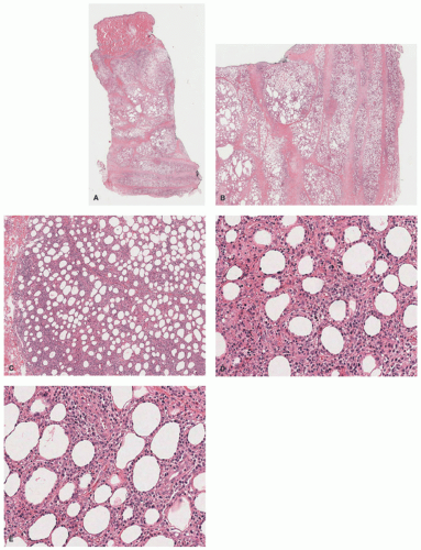
FIGURE 27-19. Subcutaneous panniculitis-like T-cell lymphoma. There is a mostly lobular panniculitis with dermal and epidermal sparing (A and B). A very dense lymphoid infiltrate in the subcutaneous lobule is seen (C). The atypical lymphocyte population shows prominent adipocyte “rimming” (D and E). Digital slides courtesy of Path Presenter.com.


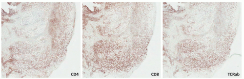
FIGURE 27-20. Subcutaneous panniculitis-like T-cell lymphoma—immunohistochemistry. The malignant infiltrate is positive for CD3 (not shown), CD8, and TCRαβ, whereas it is negative for CD4. Reprinted with permission from Gru AA, Schaffer A. Hematopathology of the skin: A clinical and pathologic approach. 1st ed. Philadelphia, PA: Wolters Kluwer; 2017.

The immunophenotype reveals CD3+, BF1+, CD8+, and cytotoxic molecules, such as TIA-1, perforin, and granzyme B. CD56 and CD30 are typically negative. Ki67 is moderately high (>50%)199 (Figure 27-20). Loss of CD2, CD5, and/or CD7 is seen in 10%, 50%, and 44% of cases, respectively.134 CD45RO is usually positive and CD45RA is negative. EBV has been rarely documented in patients of Asian descent.200,201,202,203,204

Only gold members can continue reading. Log In or Register to continue

Stay updated, free articles. Join our Telegram channel

May 8, 2019 | Posted by in Dermatology | Comments Off on Cutaneous Hematolymphoid Proliferations

Full access? Get Clinical Tree

Get Clinical Tree app for offline access