Anatomical location
Types of tumors
Suggested cryosurgical treatment
Eyelid
Benign tumors of the eyelid
(Epidermis)
Eyelid squamous papilloma
T
Eyelid seborrheic keratosis
S, P, SO
Eyelid keratoacanthoma
S, P, SO, SC, T
Eyelid cutaneous horns
T
Premalignant tumors of the eyelid epidermis
Eyelid actinic keratosis
S, SO
Eyelid sebaceous gland tumors
Eyelid sebaceous hyperplasia
P
Eyelid sweat gland tumors
Eyelid syringoma
P
Vascular tumors of the eyelid
Eyelid congenital capillary hemangioma
P
Eyelid acquired hemangioma (cherry hemangioma)
P, S
Eyelid varix
P
Eyelid lymphangioma
P
Eyelid pyogenic granuloma
P, S, T
Eyelid histiocytic, myxoid, and fibrous lesions
Xanthelasma
S
Eyelid cystic lesions simulating neoplasms
Eyelid eccrine and apocrine hidrocystoma
P
Eyelid inflammatory lesions simulating neoplasms
Eyelid molluscum contagiosum infection
P, T
Others: lid margin
(hair structure)
Trichiasis
P, S
Conjunctiva
Vascular tumors and related lesions of the conjunctiva
Conjunctival capillary hemangioma
P
Conjunctival pyogenic granuloma
P
In benign lesions, deep freezing treatments should be avoided for patients with dark skin due to the risk of hypopigmentation.
11.2 Preparation
Proper protection of the eye is mandatory. Eye protectors should be plastic or wooden, never metal. If a spoon or spatula is going to be used, any sharp, casting, or protruding element must previously be eliminated (Fig. 11.1).
Fig. 11.1
Protection of the eyelid must be made of wood or plastic (never metal)
Touching the eye surface should be avoided at all times in order to prevent epithelial damage. For lower eyelid surgery, the upper eyelid can be pushed down to the lower conjunctival fornix and kept there for extra protection of the eye. Previous eye drops of oxybuprocaine or tetracaine will be sufficient to anesthetize the conjunctival surface.
It is necessary to have a good binocular loupe or a 3D microscope [4] as well as a whole set of probes, cones, spraying tips, eye protectors, and high-quality forceps (the latter, for trichiasis/distichiasis treatment).
11.3 Tumors of the Eyelids
11.3.1 Benign Tumors of the Eyelid (Epidermis)
11.3.1.1 Eyelid Squamous Papilloma
These are also called fibroepithelial polyps, acrochordons, or skin tags. They are very common, can have different sizes, can be protruding, occur as single units or as multiples, and present in the upper and lower lids as well as on the lateral sides of the eye. Sometimes they impair the field of vision; for some patients, they are of cosmetic concern. Cryotweezers are probably the best treatment option because they are painless, multiple lesions can be treated in one session, and there is no risk of hyperpigmentation because freezing is done only to the base of the lesion and no further. This is an important consideration because skin tags are very common in 3/5 skin types (Fig. 11.2a, b). Eyelid warts are also a form of squamous papilloma. They are very common and present as solitary with a filiform/digitated shape. These are best treated by grasping them with a cryotweezer until the freezing front reaches the bottom of the lesion. One cycle is sufficient. Larger lesions can be sprayed with LN.
Fig. 11.2
(a, b) Multiple skin tags of the eyelids treated with one cycle cryotweezers (before and after treatment)
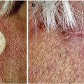
11.3.1.2 Eyelid Seborrheic Keratosis
These can be thin or thick, gray or brownish, or highly hyperpigmented, and there is usually a family tendency to present with them. They can be located anywhere in the lids (Fig. 11.3a, b) and sometimes occupy the lid margin (Fig. 11.4a, b), impairing the field of vision. Spraying with a small aperture should include a 1 mm margin of healthy skin to avoid ring-shaped recurrences or residual tumor. For lesions in the lid margin, it is best to use an appropriate size probe. The moisture in the area will help to fix the probe and control the advancing freezing zone. For those situations in which one desires to stop freezing and wants to detach the probe, a warm saline solution will do the job. One should not ever perform any forceful movements in the eyelid. The freezing with a fixed probe is best done when the patient is in a lying position.
Fig. 11.3
(a, b) Seborrheic keratosis on the lower lid margin (before and after treatment). One cycle open (spraying) technique with protection of the eye
Fig. 11.4
(a, b) Seborrheic keratosis on the upper lid margin that impaired the field of vision (before and after treatment). One cycle close (contact probe) technique
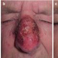
11.3.1.3 Eyelid Keratoacanthoma and Cutaneous Horns
In most dermatology reference books, keratoacanthomas are classified as well-differentiated squamous cell carcinomas. Eyelid keratoacanthomas are relatively frequent and have a typical dome-shaped appearance with a central crater or horn (Fig. 11.5). Cutaneous horns are classified by some as nonspecific keratosis. As a general recommendation, a biopsy of all cutaneous horns should be taken since some can have squamous cell carcinoma cells at the bottom. One technique that works very well for both lesions is to grab the lesion with a previously frozen cryotweezer and allow the freezing front to advance just to the bottom. Then shave the frozen block (for the histopathology study) and probe freeze the bottom of the lesion. By freezing the bottom part with a probe, any residual cells are destroyed and there will be no need for further treatment (Figs. 11.6a, b and 11.7a, b).
Fig. 11.5
Close-up look of keratoacanthoma of the eyelid
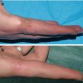
Fig. 11.6
(a, b) Keratoacanthoma on the inner angle of the lower eyelid, one freeze-thaw cycle with close (probe) technique. Before and after treatment