Upper eyelid ptosis and crease abnormalities can lead to significant cosmetic deformity. An intimate knowledge of the anatomic structures responsible for eyelid height, contour, and pretarsal show will allow precise surgical correction.
Key points
- •
Involutional changes with levator aponeurotic dehiscence is the most common cause of blepharoptosis, which presents with low margin-to-reflex distance, elevated eyelid crease, and normal levator function.
- •
Pretarsal platform is a complex anatomic structure defined by the anatomic eyelid crease and preseptal skin, fat, and brow positions.
- •
Precise surgical levator advancement can lead to appropriate marginal contour, symmetric height and allows exact eyelid crease management.
- •
Muller muscle conjunctiva resection is an effective, precise surgical procedure that requires no intraoperative patient interaction for improvement of ptosis and eyelid crease asymmetry.
- •
Eyelid crease creation and symmetry improvement can be achieved with or without ptosis repair and involves deep fixation of pretarsal anterior lamellae to the levator aponeurosis.
Introduction
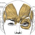
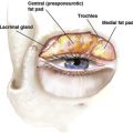
The complex anatomy of the upper eyelid is not only responsible for the upper eyelid position superior to the visual axis, but also contributes to the aesthetically pleasing lines of the eyelid margin and eyelid crease, and their relationship to the brow. The function of the upper eyelid is ocular protection and tear dispersion over the eye. Upper eyelid retractors, which are responsible for upper eyelid height and excursion, include the levator muscle and its aponeurosis, Muller muscle, and limited functions from the frontalis muscle ( Fig. 1 ). The levator muscle, which is primarily responsible for upper eyelid height and excursion, is innervated by the oculomotor nerve after its origination on the sphenoid bone in the posterior orbit. The skeletal muscle of the levator palpebrae superioris transitions into an aponeurosis after passing through the Whitnall check ligament in the superior orbit. Traditionally, the upper eyelid crease is thought to derive from the insertion of the levator aponeurosis fibers into the anterior lamellae of the eyelid. Muller muscle is an involuntary, sympathetically innervated, smooth muscle, which originates on the undersurface of the levator muscle and inserts at the superior edge of tarsus. The peripheral arcade of eyelid is an excellent landmark as it sits just above the tarsus, sandwiched between the levator aponeurosis and Muller muscle. The orbicularis muscle is a sphincter within the anterior lamellae and is responsible for eyelid protraction.