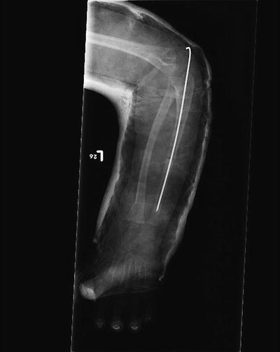
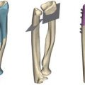
Fig. 9.1
Plain radiographs showing complete radioulnar synostosis
Classification
Wilkie, Tachdjan and Cleary and Omer have proposed various classification systems.
Wilkie [2] described two types of congenital synostosis, based on the proximal radioulnar junction. Type 1 is a complete synostosis, with the radius and ulna fused proximally for a variable distance. Type 2 is a partial synostosis involving the region just distal to the proximal radial epiphysis and is associated with radial head dislocation.
Classification according to Tachdjian’s criteria [14]:
Type 1: The radial head may be fused to the ulna or may be completely absent (known as the “headless type”)
Type 2: The radial head is malformed and often dislocated
Cleary and Omer [1] proposed a four-part radiological classification:
Type 1: Synostosis did not involve bone and was associated with an abnormal looking radial head
Type 2: A visible osseous synostosis was present, otherwise normal findings
Type 3: Osseous synostosis with hypoplastic and posteriorly dislocated radial head
Type 4: Short osseous synostosis with anteriorly dislocated radial head, which is usually mushroom shaped
Cleary and Omer Type 3 is the most common type reported in various studies. In Ramachandran et al.’s study [13], out of the six cases in their series, five forearms were classified as Cleary and Omer Type 3 (with a posteriorly dislocated radial head), while one was classified as Type 2. In the paper by Rubin et al. [15], all cases were classified as Cleary and Omer Type 3. Since there is not much functional difference between the different types and the appearances may change with time, the classifications may have not much role in deciding the management and are thus of limited clinical significance [13].
Management
Many children with forearm synostosis will not have much functional limitation and they can be treated conservatively. These children will often have mild pronation deformities less than 60°, unilateral disease and are able to compensate with radiocarpal and intercarpal wrist rotation. Often these children present to clinic when their parents or school teachers have noticed that they perform routine tasks differently from their peers.
Indication for Surgery
Most authors suggest a pronation deformity of 60° to be significant enough to merit operative intervention. In the paper by Ramachandran et al. [13] all of the patients had a mean pronation deformity of 68°. Simmons et al. [12] considered pronation of 60° as definite indication for osteotomy, while 15–60° was considered a relative indication depending on individual need of that patient. Ogino and Hikino [3] proposed that a pronation deformity of 60° created disability that needed surgery, whereas patients with a mean deformity of 20° did not complain of significant disability. These figures have varied in different studies, and some papers have considered ethnic and cultural factors that could influence decision-making.
Age at Surgery
Most children will present at school-going age. There is some variation in the literature about the age at surgery for these children. In the study by Ramachandran et al. [13], the mean age of the patients was 4.9 years (3.5–8.5 years). In the study by Rubin et al. [15], the average age at surgery was 11 years (range 9–13 years.). Hung [16] performed surgery at an average age of 6 years 3 months. There were a total of 34 patients and 52 forearms in this series. Eighteen patients (52.9 %) had bilateral deformities. They considered the ideal age for surgery to be between 3 and 6 years as it is easy to perform an osteotomy and there is significant potential for remodelling left at this age. Griffet et al. [17] considered an average age of 4–10 years as appropriate for surgery. In the study by Kanaya and Ibaraki [18] the average age was 8 years and 2 months (range 6 years 4 months to 11 years 10 months).
Operative Management
Various types of surgeries have been described. The two broad categories include either operative mobilization to restore forearm rotation or to perform an osteotomy to place the forearm in a position appropriate for day-to-day activities of the child.
Resection of the synostosis to restore forearm rotation has generally produced unsatisfactory results with subsequent loss of correction and vascular complications [18, 19]. Interposition of a free vascularized fascial flap between the separated bones has been attempted to reduce the risk of reformation of the synostosis [18, 20, 21]. Joint replacement using metallic swivel prostheses in the intramedullary canal of the radius between the supinator and pronator teres did not show good results [22].
The second group of procedures involves an osteotomy. Three types have been described to correct the deformity. The first is an osteotomy at the site of the synostosis followed by an acute correction. As the rotation here takes place over a narrow space, this may lead to excessive soft tissue tightness, loss of correction, vascular complications and neurological deficit including posterior interosseus nerve palsy [12, 23]. The second type is an osteotomy at a single site at the distal radius diaphysis [24]. The third type involves osteotomy of the diaphysis of both the radius and ulna [16, 25].
Other authors have used circular external fixators such as Illizarov to gradually correct the deformity [26]. Other techniques included osteotomies followed by derotation 10 days later [27, 28] and bone shortening by resection of bone from the synostosis [3].
Murase et al. [25] performed osteotomies in the distal third of the radius and proximal third of the ulna in patients with deformities more than 70° of pronation. They achieved good correction and only lost about 20° of correction in one case. Ramachandran et al. [13] performed a distal radius osteotomy achieving correction in all their cases (see operative technique below). Hung [16] performed a shortening by resection of 1.5 cm of bone. They measured the length of the synostosis mass in their cases and found the average length between 15 and 18 mm. Yammine et al. [29] recommended shortening the forearm by <2 cm.
Various authors have attempted separation of the synostosis with mixed results. Muira et al. [19] interposed the anconeus after synostosis separation but could not prevent recurrence with this technique. Most authors have used some sort of interposition graft after separation of the synostosis. Gill et al. [30] noted that free fat grafts worked well to prevent recurrence and performed better than Gelfoam in dogs. They also noted that pedicle fat graft was superior to free fat graft for this purpose. Langenskiold and Valle [31] demonstrated the viability of free fat grafts transplanted onto the dura up to 18 years later in four patients. Kanaya and Ibaraki [18] reported excellent results with the use of a free vascularized fascio-fat graft with no recurrence in their seven cases. They chose the lateral aspect of the ipsilateral arm as the donor site for their fascio-fat graft to ensure that surgery was confined to one limb only.
The Illizarov technique has been successfully used for this deformity. Rubin et al. [15] performed an osteotomy followed by gradual correction of deformity using the Illizarov frame achieving excellent results. They pointed out that correction should be achieved gradually as two of their patients did develop radial nerve neurapraxias when they attempted acute corrections. Bolano [26] also used the Illizarov frame but performed an immediate acute correction of 60° followed by a gradual derotation. Because of the complications encountered by Rubin et al. [15] using this technique of acute correction, they did not recommend it.
Operative Technique from Ramachandran et al. [13]
The patient is positioned supine and a well-padded tourniquet applied. An osteotomy is performed in the ulna at the mid shaft level through a subcutaneous posterior approach. A 1.8-mm Illizarov wire is passed retrogradely from the osteotomy to exit through the olecranon, and then antegradely across the osteotomy into the distal ulna. A second osteotomy is then performed in the radius at the distal diaphyseal-metaphyseal junction through a volar approach using an oscillating saw. The tourniquet is released and the forearm rotated to a position of 10° of supination. The deep fascia of the forearm is incised proximally and distally at the osteotomy sites to allow for expansion of the muscle bellies. The Illizarov wire is bent and left proud of the skin. An above-elbow plaster cast is applied with the elbow flexed to 90° and the forearm in the corrected position (Fig. 9.2). The patient is observed post-operatively for any evidence of compartment syndrome and neurovascular deficit.