Eyelid surgery consists of challenging reconstructive and cosmetic procedures. Because of the complex anatomy and corresponding vital functions of the upper and lower eyelids, the avoidance of eyelid complications is of vital importance. Complications after eyelid surgery include basic complications (infection, granuloma) and vision-threatening complications. Preoperative history, physical examination, surgical planning, and meticulous surgical technique must be undertaken to prevent complications after eyelid surgery. In addition, patient knowledge, expectations, and motivations must be determined before surgery is performed.
Key points
- •
Preoperative consultation, ophthalmologic/eyelid examination, planning, and meticulous surgical technique are paramount in avoiding postoperative complications after eyelid surgery.
- •
Counseling the patient about what they can expect in the days to weeks after eyelid surgery is extremely important.
- •
Complications can range from those that are inherent to any surgery (infection, bleeding, scar) to severe complications that are irreversible (blindness).
Introduction
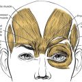
The upper and lower eyelids serve functional and cosmetic purposes. Functionally, the eyelids serve to protect the globe from injury. They are also vital to globe protection through the maintenance of tear film, distributing tear film over the cornea, and to regulate the physiologic flow of tear film. The eyelids and periorbital region are central to the perception of facial beauty and aging. The anatomy of the upper and lower eyelids is complex, and altering one structure may have consequences for the entire anatomic unit. A successful surgical procedure preserves the vital function of the eyelid, while maintaining proper symmetry and aesthetic proportions.
Cosmetic blepharoplasty is one of the most common surgical procedures performed in the United States. The main purpose of blepharoplasty is to restore a youthful appearance and bring attention back to the eyes. Eyelid reconstruction, particularly after skin cancer excision, is also common given the high frequency of skin cancer in the general population. Most postoperative eyelid complications are transient and easily treated (infection, granuloma), whereas there are select complications that can have significant consequences (ptosis, ectropion, irreversible blindness) ( Boxes 1 and 2 ). Complications resulting from eyelid surgery are prevented by detailed patient analysis, meticulous surgical technique, and appropriate postoperative care.
- •
Suture granuloma
- •
Infection
- •
Epiphora
- •
Eyelid hematoma
- •
Lagophthalmos
- •
Dry eye syndrome
- •
Eyelid retraction
- •
Chemosis
- •
Diplopia
- •
Ptosis
- •
Corneal abrasion/ulcer
- •
Retrobulbar hematoma
- •
Blindness
- •
Wound dehiscence
- •
Suture milia
- •
Suture granuloma
- •
Scar/web formation
- •
Chemosis
- •
Ptosis
- •
Deep superior sulcus
- •
Lower eyelid hollowing
- •
Eyelid crease asymmetry
- •
Overcorrection
- •
Undercorrection
Introduction
The upper and lower eyelids serve functional and cosmetic purposes. Functionally, the eyelids serve to protect the globe from injury. They are also vital to globe protection through the maintenance of tear film, distributing tear film over the cornea, and to regulate the physiologic flow of tear film. The eyelids and periorbital region are central to the perception of facial beauty and aging. The anatomy of the upper and lower eyelids is complex, and altering one structure may have consequences for the entire anatomic unit. A successful surgical procedure preserves the vital function of the eyelid, while maintaining proper symmetry and aesthetic proportions.
Cosmetic blepharoplasty is one of the most common surgical procedures performed in the United States. The main purpose of blepharoplasty is to restore a youthful appearance and bring attention back to the eyes. Eyelid reconstruction, particularly after skin cancer excision, is also common given the high frequency of skin cancer in the general population. Most postoperative eyelid complications are transient and easily treated (infection, granuloma), whereas there are select complications that can have significant consequences (ptosis, ectropion, irreversible blindness) ( Boxes 1 and 2 ). Complications resulting from eyelid surgery are prevented by detailed patient analysis, meticulous surgical technique, and appropriate postoperative care.
- •
Suture granuloma
- •
Infection
- •
Epiphora
- •
Eyelid hematoma
- •
Lagophthalmos
- •
Dry eye syndrome
- •
Eyelid retraction
- •
Chemosis
- •
Diplopia
- •
Ptosis
- •
Corneal abrasion/ulcer
- •
Retrobulbar hematoma
- •
Blindness
- •
Wound dehiscence
- •
Suture milia
- •
Suture granuloma
- •
Scar/web formation
- •
Chemosis
- •
Ptosis
- •
Deep superior sulcus
- •
Lower eyelid hollowing
- •
Eyelid crease asymmetry
- •
Overcorrection
- •
Undercorrection
Preoperative history
Before planning any surgery involving the periorbital region, a thorough patient history must occur ( Box 3 ). During this history, it is of upmost importance that patient motivations and expectations are discussed. During the history, the surgeon should look for potential “red flags” that may predispose the patient to complications in the intraoperative or postoperative period ( Box 4 ). The patient must provide a detailed list of all medications, including herbals and supplements. The use of anticoagulants and antiplatelet medications is important in that these may cause problematic bleeding, but stopping these medications in the perioperative period is a complex decision and should be done in concert with the patient’s primary care physician. In addition, the use of artificial tears regularly or on an as needed basis must be determined because many patients may not consider artificial tears as a medication.
- •
Patient expectations/motivations
- •
Ophthalmologic history
- •
Dry eye symptoms? (burning, foreign body sensation)
- •
Medications/herbals (anticoagulants/antiplatelet drugs)
- •
Does the patient use artificial tears?
- •
Systemic diseases/neuromuscular diseases
- •
Previous ocular procedures (laser in situ keratomileusis)?
- •
Previous cosmetic procedures/surgery?
- •
Relevant scarring history?
- •
Tobacco use
- •
Workplace hazards?
- •
Psychiatric/psychological difficulties
- •
Recent motivations (job/divorce)
- •
Unrealistic expectations
- •
Dry eye symptoms
- •
Thyroid orbitopathy (Graves disease)
- •
Anticoagulants/antiplatelet medication
- •
Lower lid laxity
- •
Scleral show
- •
Negative vector eyelid
In eyelid surgery, there are specific elements within the medical history that are specifically of concern. A history of other ophthalmologic conditions, dry eye symptoms, previous orbital surgery, systemic diseases, neuromuscular conditions, and scar formation should be discussed before surgery. When discussing ophthalmologic conditions or procedures, a history of laser in situ keratomileusis is important because it may predispose the patient to dry eye symptoms after surgery. Systemic diseases and neuromuscular conditions to be aware of include Graves disease, Sjögren syndrome, rheumatoid arthritis, Bell palsy, and myasthenia gravis. Scar formation is an important topic to address because certain patients, particularly those with darker skin pigmentation, may be predisposed to poor scar or keloid formation.
Periocular/ocular examination
A thorough periocular and orbital examination should be performed in any patient undergoing cosmetic or functional procedures of the eyelid ( Box 5 ). Ideally, a visual acuity measurement should be undertaken using a standard eye chart. The reactivity of the pupils and ocular motility should also be recorded. A slit-lamp evaluation is a standard examination to rule out any ocular surface irregularities and is a common examination performed by an ophthalmologist. If a surgeon does not routinely perform the previously mentioned tests, a referral to an ophthalmologist is warranted.
Ophthalmic Examination
- •
Visual acuity
- •
Pupillary examination
- •
Ocular motility
- •
Visual field testing
- •
Slit-lamp examination
- •
Retinal examination
Periocular Examination
- •
Skin color (Fitzpatrick I-VI)
- •
Brow (position, height, contour)
- •
Presence/absence ptosis
- •
Upper eyelid crease
- •
Lower eyelid fullness
- •
Negative vector lower eyelid
- •
Snap-test
- •
Distraction test (normal <6–8 mm)
- •
Schirmer test (normal 10–15 mm)
- •
Hertel exophthalmometer (normal 12–21 mm)
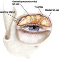
Examining and recording details about the brow and upper eyelid is essential and may help prevent complications including lagophthalmos, ptosis, upper eyelid malposition, and lacrimal gland injury. The height, contour, and position of the brow is important and this should be considered one unit, “the brow-lid continuum.” Brow and upper eyelid ptosis should be recorded and explained to the patient, in addition to any preoperative asymmetries that are noticed in the physical examination or photographs. Other important details to record are the position of the upper eye crease and upper eyelid fullness that may be a manifestation of a prolapsed lacrimal gland.
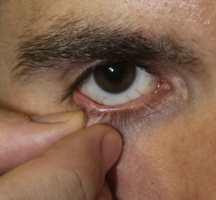
Lower eyelid evaluation is important to avoid classic postoperative complications including chemosis, ectropion, malposition of the lid, and dry eye syndrome (DES). The classic examination maneuver to assess lid laxity is the snap-test, where the examiner’s index finger is used to provide downward traction on the lower eyelid and then abruptly released ( Fig. 1 ). If the eyelid returns to the globe, this indicates a normal eyelid tone. Another examination maneuver to assess the lower eyelid is the distraction test where the examiner pulls the lower eyelid in an anterior vector. If the eyelid is able to be pulled 6 to 8 mm, this indicates lower lid laxity ( Fig. 2 ). The position of the medial and lateral canthus, the lower eyelid-cheek junction, and the presence of a negative vector eyelid should also be assessed. A negative vector eyelid is described as a prominent globe with a retrusive orbital rim ( Fig. 3 ). Patients with a negative vector lower eyelid are at an increased complication rate, particularly malposition of the eyelid.
Complications
Infection
Infection following blepharoplasty is rare given the high vascularity of the eyelids. Carter and colleagues reported a 0.2% infection rate after blepharoplasty without laser resurfacing and a 0.4% infection rate in those that had laser resurfacing. If a true postblepharoplasty infection does occur, treatment depends on the extent of disease. The most common causative pathogens are normal skin flora, but S treptococcus and Mycobacteria have also been reported. Minor cellulitis is usually successfully treated with a third-generation cephalosporin or fluoroquinolone.
Orbital cellulitis is more severe and manifests as excessive pain, eyelid swelling and erythema, decreased visual acuity, and extraocular muscle immobility. Contrast-enhanced computed tomography is necessary to rule out postseptal infection, abscess formation, or even cavernous sinus thrombosis. Streptococcus , Staphylococcus , and Mycobacterium are the most common offending bacteria that cause orbital cellulitis ( Fig. 4 ). These patients are managed with broad-spectrum intravenous antibiotics and, in cases of abscess formation, surgical drainage. Overall, the infection rate for outpatient eyelid surgery is extremely low. Perioperative prophylactic antibiotic administration is not necessary in routine blepharoplasty because topical antibiotics in the postoperative period are sufficient for infection prevention.