and Maria Siemionow2
(1)
Onep Plastic Surgery Clinic, Istanbul, Turkey
(2)
Department of Orthopaedic Surgery, University of Illinois, Chicago, IL, USA
Abstract
A new model of combined semimembranosus muscle and epigastric skin free flap was designed. The flap was based on a single pedicle consisting of the muscular branch of semimembranosus muscle and superficial epigastric vessels in continuity with femoral vessels. The anatomy of the semimembranosus muscle was studied in details. The mean length of the muscle was 38 mm and width was and 11 mm. The mean weight of the muscle was 1.03 g. The mean external diameter of the femoral artery was 1 mm and vein was 1.2 mm. Eight combined semimembranosus/epigastric skin flaps were dissected and transferred into the neck and contralateral groin regions with 100 % of success rate. This model of combined muscle and skin flap has several advantages. It is reliable, versatile, easy to dissect with long vascular pedicle and adequate vessels diameter for the anastomoses. It can be used for different applicability including: microcirculatory, pharmacological, physiological, biochemical and immunological studies.
Keywords
Semimembranosus muscle flapEpigastric skin flapCombined muscle and skin flapFree flap model in ratIntroduction
Despite numerous experimental models applied in microsurgical and transplantation research’ there is a constant need to search for new models to study different surgical phenomenons. The rat is a small animal model and as such presents with advantage of almost unrestricted availability and diversity of applications in the field of reconstructive microsurgery. Different models of free skin, muscle, myocutaneous, osseous and osteomyocutaneous flaps have been described in the rat [1–16].
A new model of combined semimembranosus muscle and epigastric skin free flap based on a single pedicle consisting of the muscular branch of semimembranosus and superficial epigastric vessels in continuity with femoral vessels was designed. The anatomy of the semimembranosus muscle was studied in details with great attention to its vascular supply. This new model of combined muscle/skin flap can be used for physiological, biochemical, and pharmacological studies, as well as for the transplantation studies.
Surgical Technique
Part I: Anatomic Dissection
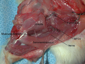
Bilateral dissections were performed in adult male Lewis rats weighing between 200 and 250 g to study the anatomy of the semimembranosus muscle, since the anatomy of the epigastric skin flap is well known (Fig. 29.1). Great attention was paid to the vascular supply of the muscle. The diameter of blood vessels and the length of the pedicle were measured. The length, width and the weight of the muscle were also measured. All measurements were performed in the same anatomical position. After completion of the dissection the animals were euthanized by overdose of sodium pentobarbital.


Fig. 29.1
Anatomic dissections of the semimembranosus muscle presenting the vascular pedicle and nerve of the muscle after the gracilis and adductor muscles were divided. MFCV medial femoral circumflex vessels [20], (with permission from Wolters Kluwer Health)
Part II: Flap Harvesting and Transfer
Combined semimembranosus/epigastric skin flaps were dissected based on the single pedicle of the femoral vessels. An elliptical skin incision was made in the right groin for harvesting of the epigastric flaps. The skin flap was elevated by including the underlying subcutaneous tissue and by preserving the superficial epigastric vessels serving as the pedicle of the flap. The superficial epigastric vessels were dissected up to their origin from the femoral vessels. A second skin incision was made extending vertically from the medial end of the groin incision down to the medial side of the knee joint. The muscular branch to the gracilis muscle and the saphenous vessels distal to the origin of the superficial epigastric vessels were ligated and divided. Proximally, the posterior gracilis muscle was separated from the underlying semimembranosus muscle. The distal part of the gracilis muscle was incised with a meticulous attention not to jeopardize the muscular branch to the semimembranosus muscle. The muscular branch to the semimembranosus muscle originating from the distal part of the femoral artery, passing through the distal part of the anterior gracilis muscle and entering the distal part of the semimembranosus muscle was identified and protected. A small cuff of the anterior gracilis muscle was left around the muscular branch for the protection. Distal to the origin of the muscular branch to the semimembranosus muscle, the continuation of the femoral vessels (popliteal vessels) and all other muscular branches were ligated and divided. Next the semimembranosus muscle was dissected from the semitendinosus muscle medially and caudofemoralis muscle laterally, and the nerve to the muscle from the tibial nerve entering the proximal portion of the muscle was identified and sacrificed. A second vascular pedicle, the medial femoral circumflex vessels taking off from the external iliac vessels and entering the proximal portion of the muscle in the form of two branches were identified and cauterized. The muscle was divided from its insertion and close to its origin and was kept attached to the femoral vessels only by its muscular branch.
Dissection of the muscular branch was continued proximally to include the superficial epigastric vessels as the pedicle of the epigastric skin flap. The superficial circumflex iliac vessels, the branches of the femoral vessels, were ligated and divided. Finally, the femoral vessels were dissected up to the level of the inguinal ligament and served as the pedicle of the flap. The combined muscle/skin flap was covered with the moist saline soaked gauze for the transfer into two different recipient sites, which included the neck region and the opposite groin of the same animal.
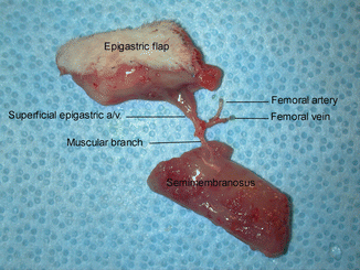
Following preparation of the vessels in the recipient sites the combined semimembranosus/epigastric flap was harvested from the donor site by dividing the femoral vessels (Fig. 29.2).


Fig. 29.2
Harvested combined semimembranosus muscle and epigastric skin flap based on the muscular branch and superficial epigastric vessels in continuation with the femoral vessels [20], (with permission from Wolters Kluwer Health)
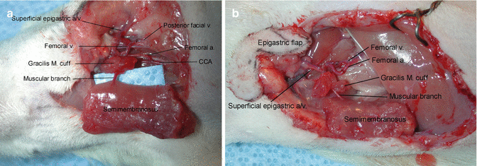
In the neck, the femoral vein of the flap was anastomosed to the posterior facial vein (end to end) and the femoral artery was anastomosed to the common carotid artery (end to side) (Fig. 29.3a). In the groin, the femoral vessels of the flap were anastomosed to the recipient femoral vessels in the end-to-end fashion (Fig. 29.3b).


Fig. 29.3
The combined semimembranosus/epigastric flap transferred into the neck region and anastomosed to the common carotid artery (end to side) and posterior facial vein (end to end). CCA common carotid artery (a) and into the contralateral groin region and anastomosed to the femoral vessels in the end-to-end fashion (b) [20]. Copyright (2005) (with permission from Wolters Kluwer Health)
The animals were anesthetized at postoperative day 7 and the transferred flaps were examined for viability by direct observation of the semimembranosus muscle and epigastric skin flaps, and the patency of the femoral vessels and the muscular branch to the semimembranosus muscle was assessed under operating microscope.


