Fig. 55.1
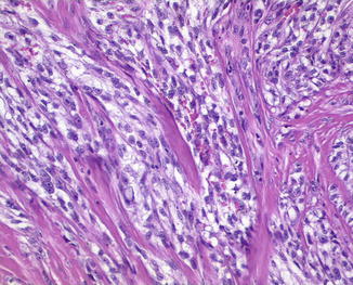
Clear cell sarcoma of soft tissue. The neoplasm is characterized by uniform nests and fascicles separated by delicate fibrous septa of hyaline collagen

Fig. 55.2
Clear cell sarcoma of soft tissue. The nests are composed of spindled or slightly epithelioid cells with finely granular eosinophilic or clear cytoplasm with large nuclei and prominent nucleoli
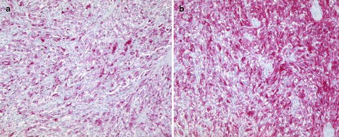
Immunohistochemical studies show that in most cases, the neoplastic cells express melanocytic markers, i.e., S-100 protein, HMB45, melan-A (Fig. 55.3a, b), and microphthalmia transcription factor (MiTF). Neural, neuroendocrine, or epithelial antigens are sometimes expressed and some tumors have melanosomes.


Fig. 55.3
Clear cell sarcoma of soft tissue. (a) The cells are positive for melan-A and (b) for HMB45
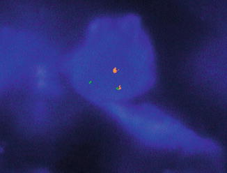
The karyotype shows a characteristic reciprocal translocation t(12;22)(q13;q12) that results in the fusion gene EWS/ATF and more rarely the translocation t(2, 22) (q34; ql2) that results in the fusion gene EWSRI/CREBI. These translocations may be identified by fluorescence in situ hybridization (FISH). Fusion type is not related to biological behavior.
Differential Diagnosis
CCS of soft tissue can easily be confused with a dermal variant of spindle cell malignant melanoma or with a metastasis of malignant melanoma because it shows the immunohistochemical profile of malignant melanoma. However, clear cell melanoma is characterized by fascicles of uniform population of tumor cells encased by delicate fibrous septa, a pattern that is seldom observed in malignant melanoma. Moreover, CCS does not display any increase in melanocytes within the epidermis or pagetoid spread of atypical melanocytes and shows multinucleated giant cells with characteristic multiple peripherally placed nuclei. Ultimately, it is characterized by a typical recurrent chromosomal translocation t(12;22)(q13;q12) that is not present in malignant melanoma and can be demonstrated by FISH analysis (Fig. 55.4) and lack melanoma-associated BRAF mutations.


