Fig. 2.1
An ulcerated vegetating tumor on the cheek of an 83-year-old woman
Pathology
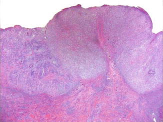
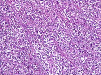
Originally, Kuo described three types of clear cell SCC, namely, keratinizing (type I), nonkeratinizing (type II), and pleomorphic (type III). Type I tumors are described as neoplasms formed by sheets or islands of clear cells with empty-appearing or “bubbled” cytoplasm with foci of keratinization and keratin pearl formation. Type II tumors are described as predominantly dermal neoplasms with parallel and anastomosing cords of cells with central nuclei and finely reticulated clear cytoplasm, without keratinization and ductal or glandular differentiation (Figs. 2.2, 2.3, 2.4, 2.5, and 2.6). In Kuo’s opinion, these could represent either recurrent SCCs or primary adnexal tumors of undetermined histogenesis. Type III tumors are described as pleomorphic neoplasms with clear cells arising from the epidermis, which show foci of squamous differentiation, dyskeratotic cells, and acantholysis and considerable perineural and vascular involvement. Kuo’s examples showed no evidence of either glycogen or mucin within the tumor cells which would support his hypothesis that clear cell changes are degenerative. However, in recent studies, examples of clear cell SCC have been shown to demonstrate glycogen accumulation in the cytoplasm of the clear cells. Only a few cases of authentic cutaneous signet-ring cell SCC have been reported. Apart from the areas with signet-ring cell appearance of the neoplastic intraepidermal or invasive cells, the tumors may show foci of conventional squamous cell carcinoma that point to the real nature of the neoplasm. The results of the studied cases indicated that mechanisms responsible for the formation of the signet-ring cells are diverse. In some cases, cytosolic accumulation of glycogen was found. In other cases, intracytoplasmic material was shown to be negative for both PAS and mucicarmine.


Fig. 2.2
The ulcerated epithelial neoplasm displays large areas of clear cells recognizable even at low magnification

Fig. 2.3


At higher magnification, these areas show parallel and anastomosing cords of cells with central nuclei and clear cytoplasm, without keratinization and ductal or glandular differentiation
Stay updated, free articles. Join our Telegram channel

Full access? Get Clinical Tree


