Chapter 6 ADIPOSE TISSUE AND THE STROMAL VASCULAR FRACTION
Adipose tissue has been considered an organ of energy storage, the largest endocrine organ, a soft tissue filler (for tissue augmentation/reconstruction), and a cosmetically unnecessary tissue discarded by liposuction. However, it is now also considered to be an important source of adult stem cells [adipose-derived stem/stromal cells (ADSCs)] and a potential tool for regenerative therapies. 1 Plastic surgeons need to learn about physiological anatomy and function of adipose tissue and its invisible cellular components [named stromal vascular fraction (SVF) after extraction] as well as how the adipose tissue and cellular components respond to surgical events such as ischemia, injury, and grafting. 2
Adipose tissue is composed of a yellow layer (mainly adipocytes) and white connective tissue. Adipocytes are densely packed in the soft yellow region, which has a capillary network. The yellow part is light because of the triglyceride in adipocytes (specific gravity 0.80 to 0.90) and thus the fat will float in water, whereas connective tissue is heavier than water (specific gravity 1.1 to 1.2).
Physiologically, adipose tissue turns over very slowly, and adipocytes have a life span of 5 to 10 years. 3 Every second, several thousand adipocytes die and are replaced with new adipocytes in our body. Adipocytes are spherical, extraordinarily large (50 to 150 µm in diameter), and filled with triglyceride. Each adipocyte has direct contact with a capillary network. 4 Adipocytes are probably the largest cells in the body; this is maintained by plasma diffusion from the capillaries. Indeed, when adipocytes exceed the maximum size in obese adipose tissue, some adipocytes will die from ischemia, and M1 macrophages will infiltrate to phagocytize the dead adipocytes. Thus obese adipose tissue is always in a state of sustained inflammation, which is considered to be the main reason for hormonal dysfunction in obese adipose tissue, as well as insulin resistance and metabolic syndrome. 5
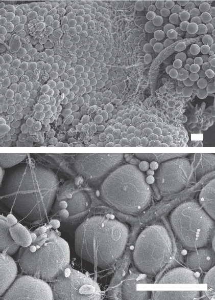
This scanning electromicroscope image of adipose tissue shows how large-sized spherical adipocytes are packed in the adipose tissue. There is significant vasculature and a capillary branch network running between adipocytes. (Scale bar = 40 µm.)
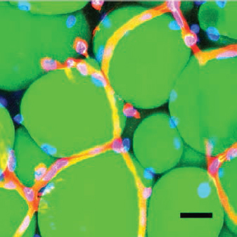
This image shows whole-mount staining of adipose tissue. Harvested, unfixed adipose tissue was stained with BODIPY (green, adipocytes), lectin (red, endothelial cells), and Hoechst 33342 (blue, nuclei). Whole-mount histologic evaluation of the living adipose tissue showed adipocytes and the capillary network. Note that every adipocyte directly attaches to a capillary (scale bar = 30 µm).
Cellular Components of Adipose Tissue
Adipose tissue has many cells other than adipocytes, although adipocytes constitute more than 90% of adipose tissue volume. Our estimate of cellular components through flow cytometry and two- and three-dimensional histologic evaluation is as follows: 1 cm 3 of adipose tissue contains 5 to 7 million cells, including 1 million adipocytes, 1 million ADSCs, 1 million VECs, and 2 to 3 million other cells (such as adipose tissue–resident macrophages and lymphocytes, pericytes, and fibroblasts). 2 , 4 , 6
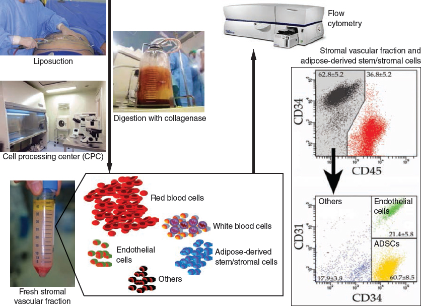
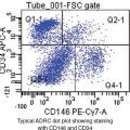
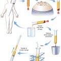
This series of images demonstrates the isolation method and cellular composition of the stromal vascular fraction (SVF) of harvested fat. SVF is isolated from lipoaspirates through collagenase digestion. SVF contains erythrocytes, CD45+ blood cells (leukocytes), CD31+/CD34− adipose-derived stem/stromal cells (ADSCs), CD31+/CD34+ endothelial cells, and CD45−/CD31−/CD34− other cells.
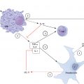
ADSCs are fibroblast-like cells that localize between adipocytes (co-localize with capillaries), in the vessel wall and in the connective tissue; most of them show perivascular localization. 7 ADSCs are considered to be progenitors of both adipocytes and vascular endothelial cells and are thus responsible for adipose tissue turnover. ADSCs are heterogeneous and are composed of monopotent progenitor cells and multipotent stem cells. 8
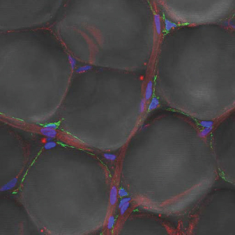
Localization of adipose-derived stem/stromal cells is shown. Whole-mount staining CD34 (green, both ADSCs and endothelial cells), lectin (red, endothelial cells), and Hoechst 33342 (blue, nuclei) reveals that ADSCs are perivascularly localized like pericytes in the capillary network, running between adipocytes.
Very recently, the existence of another type of multipotent or pluripotent stem cells (multilineage-differentiating stress-enduring [Muse] cells) in human adipose tissue, labeled by SSEA-3, was suggested 9 ; Muse cells are not localized in the connective tissue near the vessels, or are between adipocytes very sparsely. These findings suggest that there may be two types of adipose stem/progenitor cells: multipotent “stem” cells (Muse cells) located near but outside of larger vessels, and adipose “progenitor” cells (corresponding to ADSCs) located around capillaries.
Related posts:
 Chapter 7 AUTOMATED SYSTEMS FOR PROCESSING THE STROMAL VASCULAR FRACTION AND CALCULATING THE NUMBER OF STEM CELLS
 Chapter 5 ANTIINFLAMMATORY STEM CELL PRINCIPLES
 Chapter 9 GROWTH FACTORS IN THE LIPOASPIRATE
 Chapter 2 ANALYSIS OF THE PATIENT
 Chapter 10 AN OVERVIEW OF FAT GRAFTING TECHNIQUES
 Chapter 1 THE COLEMAN TECHNIQUE
Stay updated, free articles. Join our Telegram channel

Full access? Get Clinical Tree


