Chapter 5 ANTIINFLAMMATORY STEM CELL PRINCIPLES
Ongoing studies that have increased our ability to characterize fat tissue architecture have revealed its complexity. In the early 2000s it was found that fat had many different components other than adipocytes, including a population of putative stem cells. 1 These were isolated from fresh lipoaspirates in significant numbers, were able to proliferate in culture, and had multilineage differentiation potentiality. Since then, fat has been recognized and studied as a rich source of adult multipotent cells.
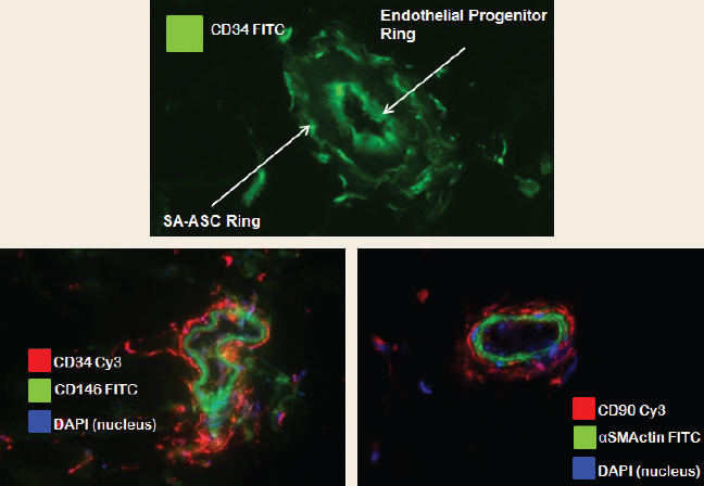
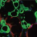
Adipose stem/progenitor populations are organized around small vessels in an anular fashion. Top: CD34+ cells form two discrete rings; endothelial progenitor cells line the lumen of small vessels, and supraadventitial adipose stromal cells (SA-ASC, preadipocytes) form an outer ring around the vessel. Left: CD146+ pericytes (green) immediately surround the vessel. Right: CD90+ cells (pericytes and SA-ASC, red) lie just beyond the adventitia, marked by alpha-smooth muscle actin (αSMA).
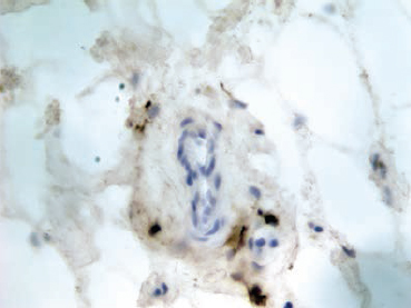
The perivascular orientation of adipose-resident leukocytes is shown. Immunohistochemical staining was performed on a Cryostat section of whole fat. Cells were stained with peroxidase-labeled anti-CD45 antibody. Antibody binding hematopoietic cells (lymphocytes and macrophages) are revealed by oxidation of 3,3?-diaminobenzidine (dark brown). Adipocytes (light brown) stain nonspecifically. The tissue is counterstained with hematoxylin and eosin, revealing the vascular cells (blue nuclei).
Associated with the microvasculature are mesenchymal and endothelial progenitor cells and their lineage-committed transit-amplifying progeny. Four distinct progenitor populations are organized around the small vessels of adipose tissue in an anular fashion. 1 Luminal endothelial progenitor cells are CD45−/CD31+/CD34+, pericytes (CD45−/CD31−/CD146+) are closely associated with αSMA cells on the surface of the vessels. Supraadventitial adipose stromal cells (CD45−/CD31−/CD146−/CD34+), sometimes called preadipocytes, surround the vessels like a sheath. The fourth population, detectable by flow cytometry but too rare to be seen by tissue staining, is a highly proliferative CD146+ CD34+ intermediate between pericytes, supraadventitial stromal cells (SA-ASC) and endothelial progenitor cells, suggesting the pericyte as a common progenitor. 2 , 3 CD45+ hematopoietic cells (T cells and macrophages) are distributed throughout adipose tissue in loose association with the vasculature. These cells have weaker intercellular adhesion and survive collagenase digestion and mechanical disaggregation far better than vascular or stromal cells. Immune cells are therefore grossly overrepresented in the stromal vascular fraction (SVF) compared with their prevalence in native adipose tissue. 4 Despite their prevalence in the SVF, the immune components and their relevance to adipose transplant biology have not been systematically addressed. In this chapter we will present new data on the T cell and macrophage components of the SVF, summarize the immunomodulatory properties of the mesenchymal component, and discuss the clinical implications of these findings.
Box 5-1 Glossary of Immunology and Stem Cell Biology Terms
Adipose-derived stromal cells (ADSC): Stem cells found in adipose tissue. Many of them can be isolated from whole or lipoaspirated fat by filtration, collagenase digestion, and centrifugation steps to obtain the cellular fraction used for regenerative medicine.
Antiglobulin: An antibody raised against another (usually cross-species) antibody. In immunohistostaining and flow cytometry, antiglobulins are often conjugated to a detection molecule such as a fluorochrome or enzyme.
APCH7: Allophycocyanin-H7 is a tandem fluorochrome used in multicolor flow cytometry. It is often conjugated directly to antibodies recognizing cellular proteins.
CD: Cluster of differentiation refers to the CD nomenclature adopted by the International Workshop and Conference on Human Leukocyte Differentiation Antigens in 1982. It is still used to classify the specificity of monoclonal antibodies. More than 330 unique CD clusters have been identified.
DAMPs: Damage-associated molecular patterns are host molecules that, when released by damaged cells, can induce an inflammatory response.
ECD: Energy-coupled dye. A proprietary tandem fluorochrome (phycoerythrin coupled to Texas Red) used in flow cytometry.
Endothelial progenitor cell (EPC): A cell capable of differentiating into mature endothelial cells that line the blood vessels.
Fc-gamma receptor: A receptor for the tail region of immunoglobulin G (called the Fc or fraction crystallizable). Fc receptors are used by macrophages and other cell types to arm themselves with antibodies, conferring the specificity of the bound antibodies.
Fluorochrome: A fluorescent dye used to label biological materials. Fluorochromes, either conjugated to monoclonal antibodies or antiglobulins, are used in flow cytometry and immunofluorescent staining to detect cellular proteins and other constituents.
HLADR: Human leukocyte antigen−antigen D related was identified as a major target of allograft rejection. Its function is to present peptide antigens to CD4+ helper T cells.
Mesenchymal stromal cells (MSCs): MSCs are multipotent stromal cells that can differentiate into a variety of cell types including adipocytes, osteoblasts, chondrocytes and myocytes.
MHC class I: Major histocompatibility complex class I molecules are expressed on the surface of almost all somatic cells. Their function is to present peptide antigens to CD8+ cytotoxic T cells.
MHC class II: The class of major histocompatibility molecules to which HLADR belongs.
PAMPs: Pathogen-associated molecular patterns are molecules associated with groups of pathogens that are recognized by cells of the innate immune system.
PECY5: Phycoerythrin-cyanine 5 is a tandem fluorochrome used in multicolor flow cytometry.
PECY5.5: Phycoerythrin-cyanine 5.5 is a tandem fluorochrome used in multicolor flow cytometry.
Pericyte: Pericytes are cells that wrap around the endothelial cells of capillaries and venules throughout the body. They are recognized as multipotential progenitor cells.
PGE2: Prostaglandin E2 is a member of the prostaglandin family. It is involved in resolution of inflammation and suppression of T cell signaling.
Stromal vascular fraction (SVF): A heterogeneous cellular fraction resulting from the disaggregation of adipose tissue into a single cell suspension.
Supraadventitial adipose stromal cells: CD45−/CD31−/CD34+ cells that form a ring above the adventitia of small blood vessels. These cells are also known as preadipocytes because they are committed to differentiate into adipocytes.
Th1: T-helper 1 cells are a subset of CD4+ T cells triggered by IL-12, IL-2. When stimulated the major cytokine that they secrete is interferon-gamma. They promote effector responses by macrophages, CD8 T cells and IgG B cells.
Th2: T-helper 2 cells are a subset of CD4+ T cells triggered by IL-4. When stimulated the major cytokines that they secrete are IL-4, IL-5, IL-9, IL-10 and IL-13. They promote effector responses by eosinophils, basophils, and mast cells.
Th17: T-helper 17 cells are a subset of CD4+ T cells distinct from Th1 and Th2 cells. When stimulated, the major cytokines secrete a host of cytokines, including interleukin 17 (IL-17). Th17 cells are localized primarily in the skin and mucosa.
Tolerogenic: Inducing immunologic tolerance, an unresponsive state to foreign antigens, particularly transplantation antigens.
Toll-like receptors (TLRs): TLRs are a class of receptor proteins that play a key role in the innate immune system. TLR are expressed in sentinel cells such as macrophages and dendritic cells that recognize structurally conserved molecules derived from microbes (PAMPs) or damaged host tissues (DAMPs).
Transit-amplifying progenitor cell: Transit-amplifying cells are a class of progenitor cells that arise from stem cells, proliferate a finite number of times, and then differentiate.
Material and Methods
IMMUNOHISTOSTAINING
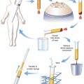
Our methods for immunostaining have been previously published. 5 Briefly, we froze small pieces of fat in liquid nitrogen. Sections were then cut on a Cryostat and slides were prepared. For immunofluorescence, sections were stained with antibodies specific for CD34, CD90, CD146 and αSMA. Nuclei were visualized by staining with the fluorescent DNA intercalator DAPI. For immunohistochemical staining, sections were stained with hematoxylin and eosin and then counterstained with antibodies to CD45 (recognizing hematopoietic cells). Cells positive for CD45 were detected by staining with an antiglobulin conjugated with the enzyme horseradish peroxidase, followed by incubation with an enzymatic substrate that stains cells brown only when they have peroxidase on their surface.
FLOW CYTOMETRY
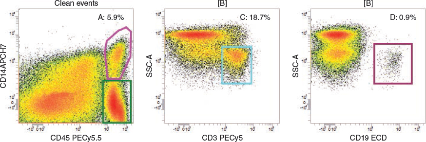
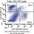
Detection of macrophages, T cells, and B cells is demonstrated in disaggregated adipose tissue. Left to right: CD45+/CD14+ events (macrophages, A gate: pink), and CD45+/CD14− events (B gate: green) are identified among “clean events.” Here clean events are defined as singlet cells with 2N to 4N DNA content, as measured by staining with DAPI (4?,6-diamidine-2?-phenylindole dihydrochloride, not shown). T cells (center) are identified as CD45+/CD14−/CD3+ events (C gate: turquoise) with low side scatter area (SSC-A). B cells (right) are identified as CD45+/CD14−/CD19+ (D gate: magenta) events with low side scatter area (SSC-A). Percentages are percent of nucleated cells (left panel), or percent of CD45 bright cells (center and right panels).
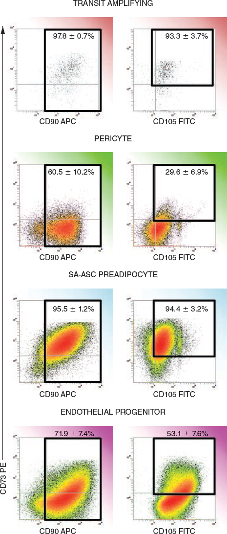
The expression of mesenchymal stem cell markers on four stem/progenitor populations of the adipose stromal vascular fraction are illustrated. Coexpression of mesenchymal-associated markers CD90, CD73, and CD105 are shown for the four stem/progenitor populations that were identified as previously reported. The percentages shown within the histogram regions display the mean ± standard error for eight independent samples studied.
Flow cytometry is a powerful single-cell technique by which a heterogeneous mixture of cells, such as SVF, is stained with multiple antibodies (each conjugated to a different fluorescent dye). The antibodies are usually directed against specific proteins of interest, many of which are identified by their “CD” number. For example, a protein that is expressed only on the surface of T lymphocytes is designated CD3. The cytometer interrogates cells one at a time and for each, measures the fluorescence intensity of each dye. In this way, quantitative measurements of the expression of multiple proteins are made on each individual cell. Although the data are multidimensional, the results (fluorescence associated with a given protein) are usually plotted in two-dimensional histograms.
Single-cell suspensions were prepared from whole fat tissue and lipoaspirates and stained as previously described. 5 , 6 Cells were first simultaneously prestained in bulk with a backbone panel containing antibodies to CD45-PECy5.5 (antibody-dye), CD3-PECy5, CD19-ECD and CD14-APCH7. Populations of interest for this report were identified by surface expression of CD45+/CD3+ (T cells), CD45+/CD19+ (B cells) and CD45+/CD14+ (tissue macrophages). Bulk-stained cells were then added to the BDT FACSCAP Lyoplate system (BD Biosciences, Mat. No. BP80394), a single 96-well plate containing fluorochrome-conjugated antibodies (three per well) covering 242 different cell-surface proteins. In this way we were able to determine the degree to which each protein was expressed on T cells, B cells, and tissue macrophages. Four independent samples were analyzed. Summarized results are given as arithmetic mean ± SEM to indicate the precision with which mean values are known, or as mean ± SD to indicate intersample heterogeneity. 7
Results
A detailed description of the hematopoietic components of the SVF has not been previously reported. To address this, we used the FACS CAP Lyoplate system to survey the cell-surface proteome of adipose resident T cells, B cells, and macrophages in SVF from the abdominal fat of four healthy subjects. CD3+ T cells represented 6.9 ± 3.5% (mean SD) of all nucleated single cells. CD14+ macrophages (7.6 ± 5.5%) and CD19+ B cells (0.2 ± 0.1%) compose the remainder of immune cells in the SVF. The high ratios of T cells to B cells (30 to 1) and macrophages to B cells (32 to 1) are far different than expected in normal peripheral blood (approximately 5:1 and 2:1, respectively), suggesting that the majority of T cells and macrophages detected are of tissue origin, and the contribution of contaminating peripheral blood is minimal.
ADIPOSE-RESIDENT T CELLS
Adipose tissue T cells and macrophage subsets are highly selected compared with those in the peripheral circulation. Adipose T cells display a unique phenotype partially resembling skin T cells, 8 especially with respect to expression of CD162, a leukocyte adhesion molecule also known as cutaneous lymphocyte-associated antigen. Like skin T cells, the majority (52.4 ± 16.3%) are CD4+, or helper T cells, the type that directly interact with MHC-class II proteins (HLA-DR) and peptide antigens presented by macrophages and dendritic cells to initiate immune effector responses. Only 5.1 ± 4.0% of the T cells are CD8+. The combination of markers usually associated with “experience” and function (naïve, memory, effector) are present in a unique combination not observed in peripheral or lymph node T cells, with the majority of cells expressing the RB isoform of CD45 (conventionally a naïve T cell marker), but in the absence of CD62L (definitively present on naïve T cells). High expression of adhesion molecules (CD44, CD11a, CD18) and markers associated with motility (CD162, CD151) is consistent with the fact that these T cells have extravasated into the adipose tissue, where they most likely participate in immune surveillance, interacting with tissue macrophages and conditioning their polarization and functionality in the process. Table 5-1 gives a more complete phenotypic description of adipose resident T cells, with the caveat that expression of single markers was determined on CD45+/CD3+ cells. This precludes definitive identification of subsets defined by the expression pattern of two or more markers. The overall profile suggests tissue resident antigen-experienced-memory or effector-memory cells. 9
ADIPOSE-RESIDENT MACROPHAGES
The great majority of adipose macrophages (92.2% ± 2.4%) express HLA-DR, the histocompatibility marker essential for presenting peptide antigens to CD4+ T cells. They are mixed concerning M1 (proinflammatory, tumor resistant, microbicidal) and M2 (antiinflammatory, matrix deposition, tissue remodeling) polarization markers. A sizable proportion of adipose macrophages express the T cell costimulatory molecule CD86 and/or the Fc-gamma receptor CD64, characteristic M1 macrophages, whereas an approximately equal proportion express CD36, CD163, and/or CD206, receptors associated with M2 macrophages. About a third express the toll-like receptor TLR-2, allowing them to respond to danger signals in the environment. Recent thinking concerning macrophage polarization emphasizes their ability to change their phenotype and function in response to cell signaling and the extracellular cytokine milieu. 10 Table 5-2 gives a more complete phenotypic description of adipose-resident macrophages, again with the caveat that they were measured one marker at a time on CD45+/CD14+ cells.
Interestingly, the nuclear receptor PPAR-gamma plays an essential role in the regulation of both adipose tissue and tissue macrophages. In adipose tissue, PPAR-gamma activation in perivascular mesenchymal stromal cells (by endogenous ligands) coordinates the expression of the genes responsible for adipose differentiation. 11 In tissue macrophages, PPAR-gamma activation occurs as part of a two-way interaction with resident Th2 T cells, which secrete the cytokines IL-4 and IL-13. PPAR-gamma activation primes tissue macrophages for M2 polarization, inducing CD36 expression in the process. 12 CD36, a scavenger receptor also known as fatty acid translocase, is involved in fatty acid uptake, 13 and is highly expressed by adipose-resident macrophages (see Table 5-1).
The complexity of the adipocyte-macrophage-T cell axis is suggested by the observation that obesity is an inflammatory condition characterized by an accumulation of tissue macrophages. 14 Not only does the number of macrophages decrease, but also their polarization shifts from M1 to M2 when the adipose burden is reduced by gastric bypass. 15 Thus it appears that in obesity, adipose is proinflammatory. This apparent conundrum is explained by the recently proposed paradigm that, like macrophages and T cells, MSCs (and presumably ADSCs) are susceptible to polarization and may promote or inhibit inflammation. 16 In addition to tissue remodeling and lipid metabolism, 17 M2-polarized macrophages have also been shown to play a role in promoting angiogenesis. 18 All of these processes are highly active in adipose tissue, which as a readily expandable and contractible source of stored energy must continually remodel and revascularize.
Taken together, adipose-resident T cells and macrophages probably play a role not only in immune surveillance, but also in regulating a milieu conducive to tissue remodeling. When concentrated in the SVF in cell-augmented lipotransfer products, they may positively contribute to the autologous adipose graft product by promoting immune surveillance after surgical intervention, facilitating wound healing, angiogenesis and graft stability, and countering scar formation.
Related posts:
 Chapter 7 AUTOMATED SYSTEMS FOR PROCESSING THE STROMAL VASCULAR FRACTION AND CALCULATING THE NUMBER OF STEM CELLS
 Chapter 9 GROWTH FACTORS IN THE LIPOASPIRATE
 Chapter 8 DETERMINATION OF FAT VIABILITY
 Chapter 2 ANALYSIS OF THE PATIENT
 Chapter 10 AN OVERVIEW OF FAT GRAFTING TECHNIQUES
 Chapter 1 THE COLEMAN TECHNIQUE
Stay updated, free articles. Join our Telegram channel

Full access? Get Clinical Tree


