Chapter 23 SIMULTANEOUS FACELIFT AND FAT GRAFTING: COMBINED LIFTING AND FILLING FOR REJUVENATION OF THE AGING FACE
The Aging Face and the Need for Fat Grafting
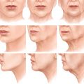
Recognizing the changes that occur in the aging face and appreciating the corresponding underlying anatomic abnormalities are essential to planning surgical procedures and recommending appropriate treatment. In most patients, problems fall into three broad categories: aging of the facial skin surface; facial tissue sagging, skin redundancy, and loss of youthful facial contour; and facial hollowing, atrophy, and/or age-related lipodystrophy. Optimal treatment will depend on the types of problems present, the patient’s priorities, and the time, trouble, and expense she or he is willing to go to to obtain the desired improvement.
Patients primarily concerned with surface aging of their facial skin may not require formal open surgical procedures and may achieve the improvement they desire through dermatologic surface treatments of the skin. Patients primarily concerned with facial sagging, skin excess, and loss of facial contour will achieve minimal if any improvement, however, if surface treatments only are employed. They will require formal surgical lifts in which sagging tissue is repositioned and redundant tissue is excised to properly address these problems and create an attractive, natural-appearing, and sustained improvement. 1 – 9
Patients with significant facial atrophy and age-related hollowing and loss of facial fat will predictably achieve suboptimal improvement from both surface treatments of facial skin and surgical lifts. Smoothing skin will not hide a drawn or hollow appearance due to loss of facial volume, and it is difficult to create natural and attractive-appearing facial contours by lifting and repositioning tissues that have abnormally thinned and that have involuted with age. Restoring lost facial volume using fat grafting is a powerful technique that represents a new paradigm for improvement of the aging face. Fat grafting is now acknowledged by most plastic surgeons and other physicians engaged in treating the aging face as the missing link in facial rejuvenation. Properly performed, the addition of fat to areas of the face that have atrophied from age or disease can produce a significant and sustained improvement in appearance that is unobtainable by other means.
WHY PERFORM A FACELIFT PLUS FAT INJECTIONS?
Why perform a facelift plus fat injections? Why not perform just the facelift? The answer to these questions lies in the multifactorial origin of facial aging, and the fact that fat is predictably lost from the face and the face becomes hollow as one ages, in addition to sagging, drooping, and other gravitational effects. A facelift procedure alone, even when performed aggressively and comprehensively, only addresses tissue ptosis, redundancy, and excess and often produces a lifted but telltale hollow, underrejuvenated look. Fat grafting, on the other hand, allows the loss of facial fat to be treated simultaneously with the facelift. All things being otherwise equal, concomitant facelift and fat injection procedures will produce a better result than either technique performed alone, and when a facelift is performed in conjunction with fat injections, loss of contour and facial atrophy can be corrected, and optimal improvement can be obtained.
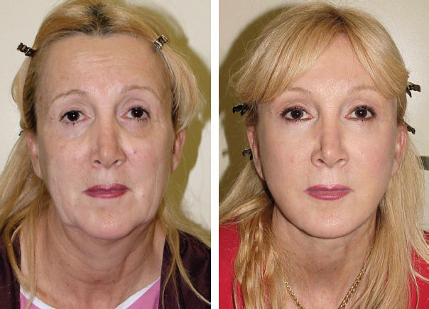
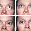
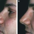
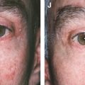
This 55-year-old patient is shown before and 1 year and 4 months after combined facelift and fat grafting. Preoperatively, it can be seen that she presented with tissue ptosis, skin redundancy, and facial atrophy. Postoperatively, she is shown after a high-SMAS facelift, neck lift, forehead lift, lower blepharocanthoplasty, and panfacial fat injections. Combined lifting and filling of her face have produced a better outcome than either technique could have alone.
*All procedures shown in this chapter were performed by Timothy Marten, MD.
VOLUMETRIC REJUVENATION, TISSUE INTEGRATION, AND THE STEM CELL EFFECT
Fat grafting has other previously unavailable advantages for surgeons who perform facelifts. Fat grafting provides volumetric rejuvenation—a new and different means by which to improve facial appearance, and a new dimension for plastic surgeons to work in. And unlike nonautologous injectable fillers, fat actually integrates with facial tissues and becomes a part of the face, promoting a more natural appearance during facial movement and producing a more sustained and long-lasting improvement. In addition, mounting scientific evidence supports the often-cited clinical observation that fat grafting may actually induce an improvement in facial tissue quality through a “stem cell effect,” and when performed with a facelift may constitute, for the first time, rejuvenation in the true sense of the word.
NOMENCLATURE
Confusion has been created for both patients and surgeons by the term stem cell facelift, a procedure in which fat is variously injected into the face with the purported but as yet unproven benefit of tissue rejuvenation mediated through a stem cell effect. In most cases, fat only is injected and a facelift is not actually performed, but in some such procedures, limited traditional facelifts are simultaneously carried out. Currently, it is a stretch of the available scientific fact to call fat grafting a “facelift,” and it is medically more correct to refer to the two procedures simply as combined but separate and distinct surgeries.
DRAWBACKS OF FAT GRAFTING
Performing fat grafting in conjunction with a facelift has certain disadvantages that must be acknowledged, including the learning curve associated with mastering any new procedure, an increase in operating room time, increased postoperative edema, a longer period of recovery, the uncertainty of graft take, and the potential for problems and complications such as asymmetries, lumps, and other irregularities. Certain patient misconceptions must also be overcome, including misguided notions held by many that injected fat can migrate or fall, or that fat grafting will make the face “look fat.”
WHY NOT JUST GRAFT FAT?
Age-related loss of facial fat rarely exists as an isolated event in healthy patients, and thus patients troubled by it can rarely be logically or appropriately treated by fat grafting alone. Isolated fat grafting is also arguably of questionable benefit to a patient with significant facial sagging and skin redundancy. Whereas aggressive filling of the sagging face with fat can yield improved contour and a smootherappearing skin surface, it generally results in an unusually large, overfilled “filler face” that appears both unnatural and unfeminine. Such an overfilled face is hard to correct in an attractive manner at a later date, and it is both more logical and practical to perform fat injections in conjunction with formal surgical lifts if needed, or after ptotic tissue has been repositioned and redundant tissue has been removed. In this way, a more natural distribution of fat is obtained and less overall fat grafting is required.
WHERE SHOULD THE FAT BE INJECTED?
Areas in need of fat grafting will vary among patients, and planning fat grafting procedures requires looking at the face in a different way—more as a sculptor and less as a tailor, as we have done in the past. Any area successfully treatable with nonautologous injectable fillers is potentially treatable with fat grafts, however, including, but not limited to, the forehead, temples, brow, glabella, radix, upper orbit (upper eyelid), lower orbit (lower eyelid), the tear trough, cheeks, midface, buccal recess, lips, perioral area, stomal angles, nasolabial fold, geniomandibular groove (GMG), jawline, chin crease, submental crease, and chin areas, and experience with fillers is a useful point of reference for planning fat additions to the face.
Perhaps the best way to decide where fat is needed is for the surgeon to study her or his previous facelift outcomes carefully and to identify areas in which the procedure has fallen short. In most cases, the most significant shortcoming for the experienced surgeon will be evident as a failure to replenish lost volume.
Opportunities to see nonsmiling photographs of patients when they were younger are highly valuable and one of the best ways to gain an appreciation of volume loss and its contribution to changes that occur as the face ages. These photos are also very helpful for educating patients as to how their face has changed with age and for explaining the need for fat grafting.
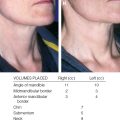
In time, and after engaging in thoughtful study of the aging face, one will gain a deeper appreciation of facial atrophy and an increasing desire to correct it. The following images show a patient before and after a facelift (and related procedures) and fat grafting, and the areas where fat was placed. It serves as a useful starting point in understanding age-related facial atrophy and how to correct it.
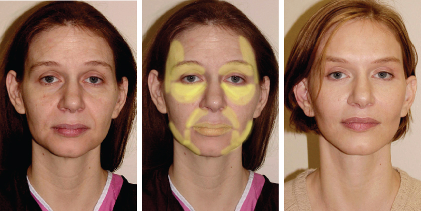
This 47-year-old patient had no prior surgery; she is shown before and after a combined facelift and fat grafting. She presented with tissue ptosis, skin redundancy, and facial atrophy. She is shown 1 year and 1 month after a high-SMAS facelift, neck lift, forehead lift, upper blepharoplasty (levator reinsertion), lower blepharoplasty, upper lip lift, and a total of 38 cc of fat injections. The shaded areas in the center photograph show where fat was placed: 1 cc was placed in each upper orbit, 3 cc was placed in each temple, 1 cc was placed in each tear trough, 3 cc was placed in each infraorbital area, 4 cc was placed in each cheek, 1 cc was placed in each nasolabial fold, 1 cc was placed in each stomal angle, 1 cc was placed in each geniomandibular groove, 3 cc was placed along each jawline, and 1 cc was placed in each lip (0.5 cc in each side). Combined lifting and filling of her face has produced a better outcome than either technique could have alone.
SEQUENCING FAT GRAFTING WITH OTHER PROCEDURES
Although there is no consensus on when fat grafting is best performed during facelift surgery, as a practical matter, it is most expedient to inject fat at the beginning of the procedure, before the facelift itself has been performed. The reasons for this include the fact that it is easier to harvest the fat at the beginning of the procedure before the face has been prepped or draped and when the patient is typically in a deeper plane of anesthesia. In the beginning of the procedure, the tissue planes of the face have also not been opened, the face is not swollen, and preoperatively made pen markings and facial landmarks are easier to identify. In addition, surgical principles suggest that it is best to limit the time the fat graft is out of the body. Perhaps the most important reason to perform fat grafting first, however, is that the surgeon is more technically and artistically energetic in the morning and will do a better job than if the procedure is performed at the end of a long facelift procedure.
Material and Methods
The technique for simultaneous facelift and fat grafting has been described previously, 1 , 3 , 10 , 11 and the principles set forth by Coleman (see Chapter 1) are observed when simultaneous facelifting and fat grafting are performed.
LOGISTICS OF SIMULTANEOUS FACELIFT AND FAT INJECTIONS
Fat grafting is often mistakenly regarded as a simple procedure that can be performed in a few minutes’ time. This is rarely the case, however, and such an attitude will lead to frustration, disruption of work-flow, and suboptimal outcomes. Fat must be harvested in a specific, atraumatic manner if viable tissue is to be obtained that can be expected to survive and take at the facial donor site. It must then be processed and injected in a technically demanding and time-consuming process. Fat grafting is also an artistically demanding activity that will require a considerable amount of the surgeon’s creative energy. When anything other than a few areas of the face are being treated, the entire procedure can encompass an hour or more, something that can overwhelm an already overburdened surgical team engaged in a long and demanding facelift operation consisting of multiple procedures. Time must therefore be carefully planned and allocated appropriately.
EQUIPMENT
Special instruments are required to harvest and inject fat, in addition to other pieces of equipment to process and organize it.
Poor outcomes are typically obtained if sharp hypodermic needles are used to harvest fat, or if fat is not processed, or if the fat is injected in a manner similar to that used to inject nonautologous facial fillers. In addition, if sharp hypodermic needles are used to inject fat (other than for a true intradermal injection), there is the potential for intravascular injection, fat embolization, and other serious problems including tissue infarction, visual impairment and blindness, and embolic stroke, and therefore their routine use is not recommended.
CHOOSING A FAT HARVEST SITE
Currently, there is no scientific consensus as to where the “best” fat for grafting should be obtained or documented evidence of one donor site being definitively superior to another. Fat harvest sites are typically chosen and marked in conjunction with the patient in such a manner as to improve her or his silhouette, although the ideal locations are arguably those areas of diet- and exercise-resistant fat collections, where the fat is biologically programmed to be stubbornly persistent throughout the patient’s life. For women, this is typically the hip, outer thigh, or abdomen, and for men it is the “love handle” and “spare tire” areas.
In thin patients, small harvests from multiple areas may be required, including the inner thighs, inner knees, upper buttocks, and anterior axilla, and the additional time this will add to the procedure must be factored into surgical planning. Preoperative assessment of the patient’s fat stores will allow proper allocation of time for the harvesting process, and appropriate pricing of the procedure.
We recommend that the harvest sites and site markings be photographed preoperatively to document what was agreed on in preoperative consultation with the patient. This helps to avoid any dispute over the preoperative condition of harvest sites after surgery.
PREOPERATIVE MARKING OF THE FACE
Facelift and fat grafting cannot be performed arbitrarily; a careful plan must be marked preoperatively with the patient seated in an upright position. Marking requires concentration on the surgeon’s part; it consumes a significant amount of time, and it is best carried out in a private area that is free from distractions. Creating an initial plan on a life-sized laser print of a photograph of the patient’s face is helpful for organizing one’s thoughts and facilitates final discussions with the patient as to which areas will be treated. In most cases, marks are made in conjunction with the patient while she or he holds a hand mirror. The goal is to create a topographic map of the deficient and atrophic areas of the face to guide the surgeon during the procedure—essentially a reverse plan of that used when liposuction is performed.
Because the aging face is undergoing a process of lipodystrophy—that is, fat is deficient in some areas and excessive in others—marks are also made where fat extraction needs to be performed when indicated. These areas can be marked in a different color or designated by some other means.
Once markings are complete, additional photographs of the marked face are taken and printed for use during the procedure, and these become part of the patient′s medical record.
INFORMED CONSENT
Some surgeons have the patient sign and date a laser print copy of the marked facial plan to avert any postoperative debates about what the patient’s preoperative requests were and what was agreed on and consented to. A companion strategy is to include language in the informed consent document stating that the discussions and designations regarding where fat is to be placed are necessarily approximate and are carried out for information and education, and that the patient consents to placement of fat during the procedure in any area that the surgeon feels, in her or his judgment, is necessary to obtain the desired effect.
ANESTHESIA
Most contemporary facelifts are time consuming and technically demanding, and the addition of fat grafting to the procedure will test the patience and composure of most surgeons. We highly recommend that the services of an anesthesiologist or other anesthesia provider be enlisted when combined procedures are performed. This is particularly important when the procedure is to be performed on a patient who is apprehensive or has a history of anesthetic difficulties, hypertension, or other significant medical problems.
Most of our facelifts are now performed with the patient under deep sedation administered by an anesthesiologist using a laryngeal mask airway (LMA). This allows the patient to be heavily sedated without compromise of the airway, but the patient need not receive muscle relaxants and can be allowed to breathe spontaneously. It is also easier to harvest fat from a heavily sedated patient, especially when fat must be harvested from multiple sites; in addition, comprehensive treatment of the face with fat is facilitated under these conditions. However, any skillfully administered anesthetic is adequate for performing the procedure.
HARVESTING FAT
Fat is harvested the day of surgery and should be harvested in a thoughtful and artistic manner that improves the patient’s figure. Thus it must typically be removed bilaterally and symmetrically, unless body contours dictate otherwise.
It is prudent to examine the patient’s body at the time of her or his consultation, unless it is obvious that plentiful donor fat is present. Thin patients with limited fat stores or patients who have undergone prior liposuction often present significant challenges when harvesting fat, and extra time and effort will be required to obtain fat from them. Anesthesia and operating room times and the surgeon’s fee must be calculated accordingly.
Fat is harvested after anesthesia is initiated, but before prep of the face. In all but the unusual case, a complete prep of the torso is not necessary. Typically, a limited prep of the marked area is made and a sterile field is established about the prepped area.
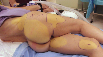
If fat is to be harvested from the hip and/or lateral thigh, as is usually preferred, the patient is turned to a semilateral decubitus position; prep, drape, and harvest are performed, and the patient is then turned to the opposite side, where the harvest procedure is repeated. If the patient is positioned carefully, this technique can be used to simultaneously harvest fat from multiple additional sites, including the inner knee, posterior inner thigh, outer thigh, upper buttocks, hip, waist, and flank area. Obtaining fat from multiple sites is particularly important in thin patients with minimal fat stores.
Areas from which fat is to be harvested are conservatively infiltrated with 0.1% lidocaine with 1:1,000,000 epinephrine solution using a specially designed multiholed local anesthetic infiltration cannula, allowing adequate time for a proper anesthetic and hemostatic effect. Approximately 1 cc of this dilute local anesthetic solution is injected for every 3 cc of anticipated fat removal. It is not necessary or desirable to infiltrate in a tumescent fashion, because overwetting the donor site will result in an overdilute harvest and more time spent in the harvesting process. Local anesthetic should be injected, even if deep sedation or general anesthetic is used, to limit the overall amount of inhalation anesthetic and narcotics used.
Fat is then harvested with a special harvesting cannula attached to a 10 cc syringe using gently applied syringe suction to avoid vacuum barotrauma to the tissue. Fat harvested with these cannulas easily passes through injection cannulas as small as 0.7 mm. If fat is harvested with too much applied suction, poor graft survival and compromised outcomes are possible.
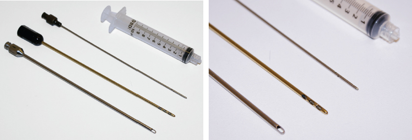
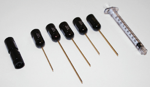
On the left, shown from the top down, are a 10 cc Luer-Lok syringe, a 1.9 mm local anesthetic infiltration cannula, a 2.4 mm Tulip Tri-port harvesting cannula (a similar Caraway harvester is also frequently used), and a 2.4 mm Coleman harvesting cannula. A closeup of the instruments’ tips is shown on the right.
In general, at least twice as much fat is harvested as is anticipated will be used to ensure that an adequate supply of processed fat will be available for use on the face. Even larger amounts are harvested if the surgeon wishes to use more high-density stem cell–rich fat (see discussion that follows).
Once fat harvest is complete, the stab incision used to obtain the fat is closed with a simple interrupted suture of 6-0 nylon. The harvest site is then washed free of prep solution, and the sutured site is covered with a TegaDerm dressing.
PROCESSING HARVESTED FAT
Harvested fat is almost never uniform in character and concentration as extracted from donor sites, since each syringe will contain a variable amount of fat, blood, local anesthetic, and ruptured fat cells (“oil”), and some type of processing is necessary to consistently obtain uniform material for injection. Although fat can be separated from the oil and water fractions using a “tea strainer” type of sieve or rolling it on Telfa gauze, the majority of “growth factors” and “cellular messengers” are likely lost when this is done. In addition, it is now known that not all fat cells are the same and that the “high-density adipocytes” are the most stem cell rich, and processing fat through a sieve or by rolling it on Telfa gauze does not provide a means of concentrating these.
Centrifugation, as advocated by Coleman, conversely allows separation of the oil (ruptured fat cells) and water (blood and local anesthetic) fractions from the fat cells, while simultaneously concentrating these other potentially important components, and has been our favored method of fat processing for almost two decades. In addition, centrifugation concentrates the high-density adipocytes and allows the surgeon to use this “supercharged” fat, which is thought to be superior in quality and higher in “stem cells” for grafting into critical areas (such as the orbits and lips), or even the entire face if an adequate overharvest of fat is done.
Before centrifugation is begun, a sterile, disposable plastic Luer-Lok cap is placed on the end of the syringe to keep harvested fat inside it, and the syringe plunger is removed from the syringe barrel. Capped syringe barrels containing unprocessed fat are then loaded into the centrifuge rotor in a balanced fashion and centrifuged for 1 to 3 minutes at 1000 rpm. Most small, portable centrifuges available for this purpose are inexpensive and have rotors or rotor syringe sleeves that can be sterilized so that the fatcontaining syringe barrels remain sterile and can be handled by the scrubbed surgical team after centrifugation.
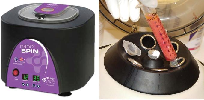
A small, portable countertop centrifuge typical of that used for operating room processing of fat is shown on the left. On the right is a closeup view of a centrifuge rotor being loaded with unprocessed fat in 10 cc syringes. Note that the tip of the syringe barrel has been sealed with a disposable plastic cap. The removable and sterilizable metal sleeves shown fit into the rotor to keep syringe barrels containing fat sterile and allow them to be handled in the sterile field after centrifugation. Other centrifuges are designed to allow the entire rotor to be sterilized.
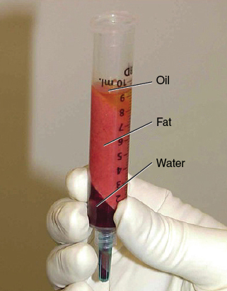
Once centrifuged, syringe barrels containing spun fat are removed; they will be seen to contain an upper oil, central fat, and a lower water (blood and anesthetic solution) fraction.
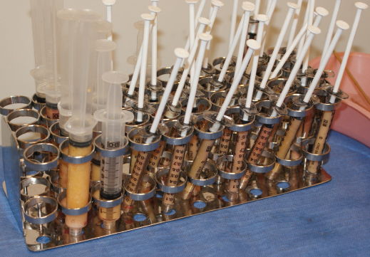
The typically blood-tinged water/local anesthetic component is separated by simply removing the syringe tip cap and allowing it to run out, and the cap is then replaced. The oil fraction is then poured off out of the top of the syringe. Telfa sponges can also placed inside the syringe barrel to wick up the small amount of residual oil present after the majority has been poured off. If fat overharvest is done, this wicking of residual oil with Telfa sponges need not be performed, and the top 1 or 2 cc of fat from each syringe is simply discarded. A test tube type of rack to hold the syringes containing fat greatly facilitates fat-processing activities. The rack also conveniently holds 1 cc syringes, syringe components, and other equipment used in the fat grafting procedure.
The bottom 2 cc of fat present in the syringe after centrifugation consists of stem cell–rich high-density adipocytes (or adipose-derived stromal cells [ADSC]) and this fat can be segregated and used preferentially in the procedure and for critical areas such as the orbits, lips, and tear trough area. If adequate donor fat is available and overharvest is done, enough high-density fat can be obtained that the entire facial fat grafting procedure can be performed using it. Such a strategy is currently regarded by many surgeons as one that holds the most potential for the best stem cell effect. In patients with limited fat stores, unused fat can be reinjected into the buttocks or a like area, essentially to “bank” it for possible future use.
FAT AND PLATELET-RICH PLASMA
A number of surgeons have advocated mixing harvested fat with platelet-rich plasma (PRP), asserting that this enhances graft take and improves overall outcomes. Mounting evidence suggests that this is not true, however, and currently fat grafting is best thought of as an art project, not a science experiment.
PATIENTS WITH PREVIOUS FILLER USE
Patients who have previously received filler injections present a specific challenge to the surgeon performing fat grafting and are arguably suboptimal candidates for the procedure. Often more filler is present than patients are aware of or admit to, and the presence of filler hides the extent of the actual deformity. The presence of residual filler will also likely compromise graft survival, make a uniform take less likely, and compromise the ultimate outcome of the procedure. Experience has shown that even fillers placed several years previously can make procedures more difficult and the outcomes less predictable.
Filler patients are a moving target and are difficult to assess and treat. Patients who have undergone HA filler treatments are more likely to have suboptimal graft survival, uneven take, and may require multiple staged treatments. HA-based fillers can be dissolved with hyaluronidase (Vitrase, Wydase), and it is recommended that HA filler be removed a few days or more before treatment. If filler is removed at the time of the procedure, the patient does not have the opportunity to see what he or she really looks like without the filler, and the extent of the problem is not evident to the surgeon. When HA filler is dissolved with hyaluronidase, the patient will also have residual inflammation and is still likely to be a suboptimal candidate for fat grafting.
Patients presently using or having used non-HA–based fillers such as silicone or polymethylmethacrylate (PMMA) often present an even more perplexing problem. Present use of poly-L-lactic acid (PLLA) or previous PLLA filler use can be particularly problematic because of chronic inflammation, internal fibrosis, and related tissue compromise. PLLA users should be informed that they are more likely to have suboptimal fat graft survival, uneven fat take, and will need multiple staged fat graft treatments.
INJECTING FAT
After centrifugation and the oil and water fractions have been discarded (and high-density fat segregated, if desired), the fat is gently transferred into 1 cc Luer-Lok syringes using a transfer coupling, because proper injection in very small aliquots cannot be made with a 10 cc, 5 cc, or even a 3 cc syringe.
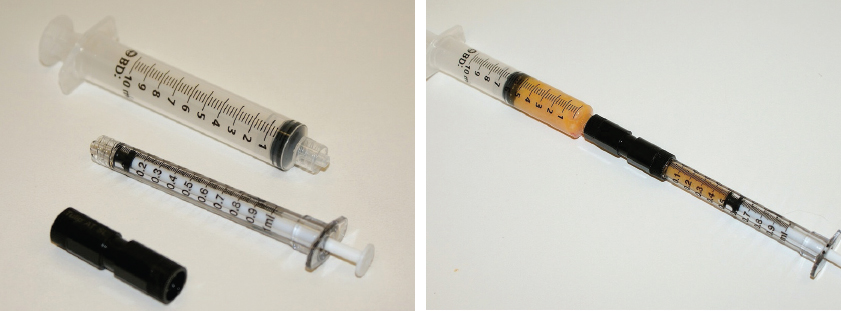
A 10 cc Luer-Lok syringe, 1 cc Luer-Lok syringe, and Luer-to-Luer transfer coupling are shown.
Nerve blocks are then performed with a 0.25% bupivacaine with epinephrine 1:200,000 local anesthetic solution, and adequate time is allowed for a complete anesthetic and hemostatic effect. It is typically not necessary to directly infiltrate areas to be treated with local anesthetic if nerve blocks are performed correctly and sedation is administered.
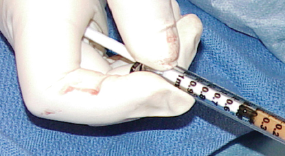
Next, 0.7 mm, 0.9 mm, and 1.2 mm (22- to 18-gauge) cannulas are used to infiltrate fat into the face through small stab incisions made in the facial skin made with a No. 11 blade scalpel or a 20-gauge needle. These incisions are so small that they will not require suturing on completion of the procedure.
Infiltration is made in multiple passes, injecting fat on both the “in” and “out” strokes in planes appropriate for the area being treated, usually from two separate injection sites, and feathering into adjacent areas. Injecting from two separate injection sites allows crisscrossing of cannula passes during graft placement, provides smoother fat infiltration, and helps avert a “row-of-corn effect” that may result if injection is done from only one site.
How Much Fat Should Be Injected? Is Overcorrection Necessary?
Deciding how much fat needs to be injected at a given site requires empirical information, and the surgeon cannot simply rely on what she or he sees in the operating room. In general, there is a tendency to treat most areas too conservatively if amounts to be injected are determined by intuition alone, and some overcorrection is needed, because not all of the grafted tissue will survive. Additionally, more fat will be needed than one would use to fill a similar defect with a nonautologous filler. In most cases, the amount needed exceeds what intuition and direct observation suggest; the amount is best decided preoperatively, based on the degree of deficiency seen in the patient’s preoperative photographs.
As a practical matter, the degree of deficiency in each area of the face can be rated as small, medium, or large, and there is an empirical range of volume needed for the typical treatment of each area that is then chosen and applied. These empirical ranges have been previously published by Coleman (see Chapter 1), Marten, Marten and Elyassnia, and other surgeons 1 – 3 , 9 ; the current range recommendations appear throughout this chapter in the sections that address specific areas of the face. For small, medium, and large problems, the volumes chosen will be on the low, midrange, or high end of the empirical volume range, respectively. For men and for women with larger faces, the recommended amounts should be increased by 25% to 35%.
How Is the Fat Injected?
As the cannula is advanced in the tissue, the surgeon should feel for resistance, and if resistance is felt, a small injection is made. Approximately 0.05 cc or less should be injected per pass. This corresponds to 20 to 40 back-and-forth passes or more for each 1 cc syringe of processed fat. If tissue resistance is not felt as the injection cannula is advanced, this indicates that a pass and injection has likely already been made in that area, and no injection is placed there; the cannula is directed to another area. The goal is to inject the fat in a way that optimizes its chance of developing a blood supply and surviving. The surgeon’s mental model should be one of scattering tiny particles of fat into the recipient site in the target plane in multiple crisscrossing fine trails so that each particle sits in its own compartment and has maximal surface contact with perfused recipient site tissue. If fat is injected in a bolus, fat cells will be clumped together, and only those on the periphery of the injected area will have tissue contact and will be likely to survive. Most of the more centrally situated fat particles will only have contact with each other, will be less likely to survive, and can lead to the formation of oil cysts. Put in more practical terms, the procedure should be thought of as analogous to “spray painting,” not “caulking.”
Advancement and withdrawal of the injection cannula will typically be made slowly by beginning injectors, but as familiarity with the technique is acquired, the movements can and should be made faster. Ultimately, all other things being equal, faster movements are desirable in that if the injection cannula is constantly in motion, intravascular injection is less likely, and the likelihood is reduced that an accidental bolus injection into one area will be made. Rapid back-and-forth movements also ensure the smoothest and most uniform “spray painting” and infiltration of fat at the same time.
How the syringe is held is also important to avoid overinjection and to control the volume expressed from the cannula with each pass. If the syringe is held in the manner one would traditionally use to give an injection with the thumb on the end of the syringe plunger, it is easy to inject too much fat if tissue resistance changes or injection cannula resistance suddenly decreases. More control can be generally be maintained, and overinjection more easily avoided, if the syringe is held with the end of the plunger in the palm of the hand. Although a bit of practice is required at first, when the syringe is held in this manner, a slight closing of the hand results in only a small amount of fat being expressed from it, and overinjection of any one area can more readily be avoided.
Although cannula obstruction is uncommon if fat is harvested and processed as described, if a cannula becomes blocked, additional injection pressure should not be applied, because this is the most common cause of a sudden and unintentional overinjection. It is better in such circumstances to simply withdraw the blocked cannula, pass it to the surgical assistant, and continue with a different one. The assistant can then clear the obstruction while the surgeon continues to work. Typically, the cause of the obstruction will be a particle of fat or subcutaneous debris at the interface of the cannula and the inside of the cannula hub, which is most easily cleared by removing the cannula from the syringe and extracting the syringe and extracting the particle of fat or fragment tissue debris from inside the back of the hub with a fine forceps.
How Deep Should the Fat Be Injected? In What Layers Should Fat Be Placed?
Injections will necessarily be made in different planes, depending on the areas being treated. In many areas where multiple tissue layers are present and the overlying skin is thick, injection can be made comprehensively at the treated site from the periosteum to the subdermal layer. These areas typically include the GMG, piriform aperture, midface, cheek, and the chin. In other areas, injections must be placed more specifically because of the anatomic characteristics of the treated sites and to avoid creating irregularities. These areas include the temples, upper orbit, lower orbit, tear trough, lips, jawline, and the dorsum of the hands. The easiest areas for the beginning injector to treat are the areas in the former category. Initially it is wise to stay deep and place most of the graft in a predominantly preperiosteal plane. Once the surgeon gains familiarity with the technique, areas in the latter category can then cautiously be treated; however, these will require very careful and skilled placement of fat.
Geniomandibular (Prejowl) Groove
Injection of the prejowl/GMG area with fat has a high aesthetic payoff and is a good area for the beginning injector to gain experience with the technique. Although not immediately intuitively obvious, filling the GMG creates a strong, uninterrupted aesthetic line from chin to the posterior mandible and is a highly desirable improvement on both the male and female face. The effect is similar to that when a Mittleman prejowl implant is placed, but fat grafting provides the advantages of an autologous procedure and is arguably simpler to perform.
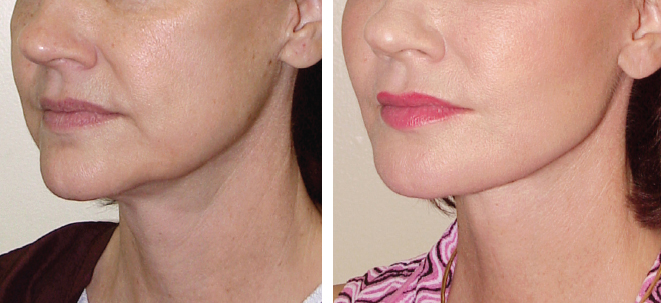
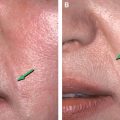
This patient is shown before and after a facelift and fat grafting; she presented with deep geniomandibular grooves. Preoperatively, her chin appears narrow and pointed, and there is poor continuity between the chin and jawline. Fat grafting was performed to fill the GMG (prejowl sulcus) area. A chin implant was not placed. Postoperatively, her chin appears broader and more aesthetically integrated with the jawline. Fat was also used to strengthen the posterior jawline and lower the mandibular angle and to fill the lips, nasolabial fold, cheeks, and infraorbital areas.
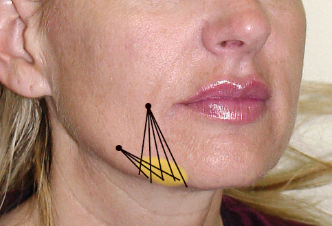
Incision sites and the plan for injecting fat into the prejowl/GMG areas are shown. Fat grafting of this area is performed with a 4 cm long, 0.7 mm diameter (22-gauge) cannula, and fat is placed in all tissue layers between the periosteum and skin. Typically, 1 to 3 cc of fat is placed on each side. Fat grafting of the geniomandibular area has a low level of difficulty.
Related posts:
 Chapter 22 A MODEL OF AGING: A PARADIGM CHANGE
 Chapter 29 STRUCTURAL FAT GRAFTING IN THE NECK
 Chapter 25 STRUCTURAL FAT GRAFTING IN THE NOSE
 Chapter 26 STRUCTURAL FAT GRAFTING IN THE NASOLABIAL FOLD
 Chapter 28 STRUCTURAL FAT GRAFTING IN THE CHIN AND JAWLINE
 Chapter 24 STRUCTURAL FAT GRAFTING IN THE SUPRAORBITAL AREA
Stay updated, free articles. Join our Telegram channel

Full access? Get Clinical Tree


