Case 7 Malignant Skin Lesion
7.1 Description
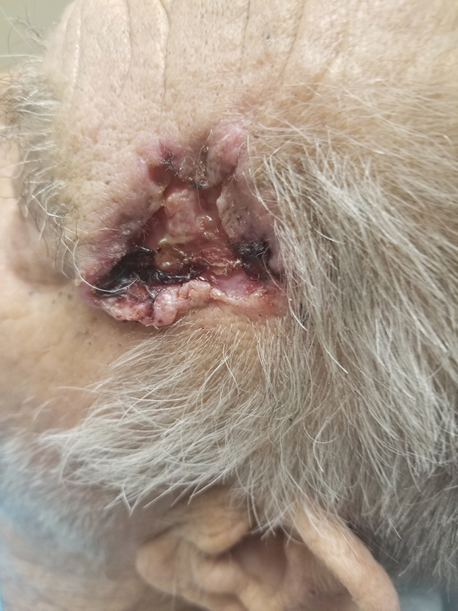
Large, eroded, pearly pink plaque with telangiectasia located on the left temple, involving the temporal hairline, and demonstrating evidence of ulceration.
There is significant concern for malignancy.
7.2 Work-Up
7.2.1 History
Length of time the lesion has been present
Associated symptoms: Pain, scabbing, itching, bleeding, and hyperkeratosis
Sun exposure history, tanning bed use
History of facial surgery, especially surrounding the nose
Previous surgery may affect reconstructive options
Primary tumor versus recurrent tumor
Previous treatment with cryotherapy, topical medications, electrodesiccation and/or curettage
Scar in area of malignancy
Complicating comorbidities: Cardiopulmonary/peripheral vascular disease, diabetes, tobacco product use, steroid use, anticoagulation, and chemotherapy
Supplementation with vitamin E, fish oil, krill oil, omega, and garlic
History of radiation, immunosuppression (organ transplant recipient, AIDS)
Personal or family history of skin cancer
Genetic conditions: Xerodermapigmentosum, Gorlin’s (nevoid basal cell) syndrome, albinism
7.2.2 Physical Examination
Detailed evaluation of affected area and surrounding face/neck to assess the lesion with or without dermatoscopic analysis/magnification
Characteristics of affected area (e.g., hair bearing, adjacent skin laxity, and pigmentation)
Skin lesion findings
Size, color, shape of lesion, skin irregularity, hyperkeratosis, and ulceration
Confirm absence of involvement of deeper structures (e.g., parotid and facial nerve)
Lymph node examination to assess for signs of metastatic disease
Full body integument examination
7.2.3 Diagnostic Studies
Establish a diagnosis: If patient presents without previous treatment, a biopsy should be performed.
Full-thickness incisional versus excisional biopsies may be performed
If the concern is for an atypical pigmented lesion, an excisional or shave biopsy should be performed to evaluate as much of the lesion as possible to minimize sampling error
Avoid shave biopsies of a portion of a pigmented lesion as they may lead to incomplete assessment of the lesion, particularly in melanoma, where the depth of a tumor is critical to prognosis
Shave biopsyis an acceptable method of assessment if the concern is for a nonmelanoma skin cancer (basal or squamous cell carcinomas)
Imaging may be necessary after an initial diagnosis is established, especially if deeper tissue invasion is a concern or in lesions of the scalp where one is suspicious of possible bony penetration
Magnetic resonance imaging (MRI): Useful adjunct to determine extent of tumor and lymph node status in cases of aggressive tumor histology (e.g., perineural invasion)
Related posts:


Stay updated, free articles. Join our Telegram channel

Full access? Get Clinical Tree


