Case 6 Pediatric Mandible Fractures
6.1 Description
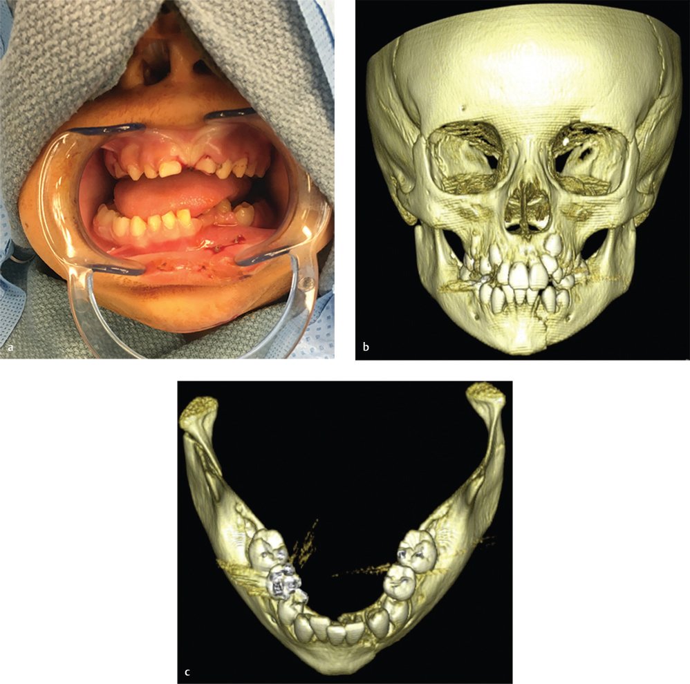
Clinical photo reveals intraoral injury including dental injury and crossbite of the mandible with evidence of mandibular widening
Computed tomography (CT) scan reveals left parasymphyseal fracture with mild displacement
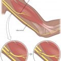
Bilateral subcondylar fractures: right side with loss of vertical height and left with minimal angulation without loss of height
6.2 Work-Up
6.2.1 History and Physical Examination
Complete trauma evaluation
Evaluate airway, breathing, and circulation
Rarely, intubation may be necessary for airway protection
Cervical spine evaluation
Assessment of associated injuries
History and examination are more challenging in children due to limited maturity and inability to articulate subjective complaints
May obtain history from parent or guardian
Beware of child abuse/neglect. Concern for these issues must be reported to child protective services.
Inspect face for asymmetry, areas of tenderness, swelling, or ecchymosis
Occlusion may be difficult to determine in younger patients as teeth are widely spaced and naturally mobile
Chin laceration may indicate superiorly directed force consistent with condylar fractures
Deviation of jaw opening or limited mobility
Intraoral examination to evaluate for lacerations or hematomas
State of dentition
Children aged 6–12 years will present with various states of mixed dentition. Younger children will have permanent tooth roots deep to their primary dentition. These factors will critically influence a surgeon’s options for reconstruction of injuries.
Presence of mixed dentition with teeth in varying stages of eruption makes evaluation of malocclusion challenging in pediatric patients.
Assess for fracture, stability, tooth root exposure, and dental caries.
6.2.2 Pertinent Imaging or Diagnostic Studies
High-resolution maxillofacial CT: Gold standard for evaluation of facial trauma. Three-dimensional reconstructions may assist in evaluating injury. It may require sedation in young children.
Panorex: Panorex requires patient’s cooperation and the patient must be upright for the study. It permits visualization of entire mandible and localization of developing permanent dentition. Towne’s view is used to improve evaluation of condyles, if necessary.
Plain radiography (Mandible Series: anteroposterior (AP), lateral, oblique, and open mouth Towne’s views): This study is of limited benefit in younger patients whose skeletons are less calcified than adults.
Related posts:


Stay updated, free articles. Join our Telegram channel

Full access? Get Clinical Tree


