11
Cardiothoracic transplantation
Introduction
The first successful heart transplantation undertaken on December 1967, at least in the public eye, represents a defining moment in surgical treatment of heart diseases in the 20th century. Since then heart transplantation has evolved from an experimental procedure to an effective therapeutic strategy for end-stage heart disease. In the current era the median survival or half-life (the time at which 50% of transplant recipients remain alive) is 11 years. For adult and paediatric patients surviving to 1 year after transplant, the median survival has reached 14 years. Many hundreds of patients have now lived past 25 years since their transplant procedure.1
Nevertheless, the success of heart transplantation has raised expectations that under present circumstances it cannot fulfil. On one hand, due to improved management of ischaemic heart disease and increased longevity, the number of patients with heart failure is growing.2 On the other hand, there is a decrease in the number of cardiac transplantations due to donor organ constraints. This disparity between the number of donors and potential recipients has stimulated research to find new alternatives to transplantation. However, these have so far had little impact on the current practice of heart transplantation. The advent of novel therapeutics and surgical options for impaired ventricles may in selected patients defer consideration for transplantation, and clinical guidelines have been provided for this purpose.3 The contemporary practice of heart transplantation with respect to indications, surgical techniques, and donor and recipient management will now be reviewed.
Indications for heart transplantation
The reason for undertaking heart transplantation is to prolong life and to improve its quality. The indications for adult heart transplantation have remained essentially unchanged over the last three decades and at present are predominantly coronary artery disease-related ischaemic cardiomyopathy (38%) and non-ischaemic cardiomyopathies (53%). Adult congenital heart disease (3%), valvular heart disease (3%), repeat transplantation (2.6%) and miscellaneous diagnoses (1%) constitute the rest.1
The indications for paediatric heart transplantation (< 18 years) are different to adults. In our series of 182 paediatric heart transplants (1987–2009), 64% were undertaken for cardiomyopathy and 36% for congenital heart disease.4
Aetiology of heart disease
End-stage heart failure has become a major medical problem.2 The increasing prevalence with the rising age of the general population in most societies accounts for a large proportion of healthcare spending due to frequent hospital admissions.5 In the aetiology of congestive heart failure (CHF), we differentiate primarily ischaemic from other cardiomyopathies and congenital heart disease during transplant candidate assessment.
Ischaemic heart disease
This constitutes the largest group requiring heart transplantation. These patients can present in a variety of ways, from being acutely ill after myocardial infarction on mechanical support to being chronically ill with heart failure with or without previous surgical or catheter-based intervention. Unfortunately, there are no conclusive prospective studies comparing conventional treatment methods with heart transplantation to provide guidance in risk–benefit assessment. A digest of current thinking would indicate that heart transplantation would definitely be indicated in a patient with severe heart failure with poor ventricular function (ejection fraction < 15%), symptoms of heart failure with little or no angina, diffuse coronary artery disease, absence of reversible ischaemia and/or poor right ventricular function (ejection fraction < 35%). What is clear is that patients with ischaemic cardiomyopathy who develop heart failure are likely to have a worse prognosis than non-ischaemic patients.6
Non-ischaemic cardiomyopathy
Certain types of cardiomyopathies can show reversibility, and a period of observation with medical treatment should be tried before listing. These include lymphocytic myocarditis, peripartum cardiomyopathy, hypertensive cardiomyopathy and alcoholic cardiomyopathy.7
Indications for paediatric patients are similar; however, the risk of death is highest during the first 3 months after presentation, therefore failure of aggressive medical treatment early in the course of the disease should lead to early assessment for transplantation. Acute myocarditis needs a special mention as the finding of acute inflammation on biopsy is a favourable prognostic sign for subsequent recovery.8
Congenital heart disease
An increasing number of patients with congenital heart disease and heart failure are now presenting in adulthood. This group is particularly challenging due to multiple comorbidities. These include previous complex surgeries, human leucocyte antigen (HLA) sensitisation, and presence of profound cyanosis and erythrocytosis.9
Recipient evaluation and selection
Patients are evaluated for transplantation once a referral has been made. We admit the patient for a few days for assessment. During this period there is a systematic evaluation of both the physical and psychological state of the patient; it also gives an opportunity to develop a rapport between the patient, relatives and the multidisciplinary team. The protocol used in our own centre for assessment is summarised in Box 11.1. The assessment process is designed to answer the following questions:
1. Does the patient fulfil the selection criteria for heart transplantation?
2. Are there any contraindications to transplantation?
Selection criteria
The limitation of this technique is that it can be influenced by body composition, individual motivation or general deconditioning. Some centres have incorporated the heart failure survival score (HFSS) to their preoperative assessment. The score consists of seven variables – resting heart rate, left ventricular ejection fraction, mean arterial blood pressure, interventricular conduction delay, peak exercise oxygen consumption (VO2), serum sodium and ischaemic cardiomyopathy. Using these variables Aaronson et al. developed a mathematical model to predict outcome with medical management.10 This score, along with maximal oxygen consumption (VO2max) and clinical assessment, can bring some rigour to the selection process for transplantation.
Contraindications
Contraindications to heart transplantation are summarised in Box 11.2. These can be classed in three groups:
1. Factors that increase perioperative mortality, e.g. elevated pulmonary vascular resistance.
2. Factors affecting long-term prognosis.
Pulmonary vascular resistance (PVR) of more than 6 Wood units has been considered an absolute contraindication to heart transplantation but with the introduction of nitric oxide, use of a bicaval anastomotic technique, early implantation of ventricular assist devices, and increasing use of perioperative phosphodiesterase inhibitors and isoprenaline, good results can be obtained in patients who formerly would not have been offered the opportunity of transplantation. Nevertheless, the presence of an elevated PVR should not be taken lightly as the donor right ventricle generally tolerates a systolic pressure of more than 50 mmHg poorly and would acutely fail. In our own practice a PVR > 3 Wood units would be considered a relative contraindication to transplantation. The transpulmonary gradient (TPG) (see Box 11.3) represents the pressure gradient across the pulmonary vascular bed and is independent of the pulmonary blood flow. Some consider the elevation of this above 14 mmHg as a more useful indication of significantly raised PVR as this is independent of the cardiac output, which may be poor in these patients. We rely more on this criterion and generally consider a fixed TPG of 12 mmHg and above as an absolute contraindication. In paediatric patients a higher TPG can be considered as it could be overcome with a larger sized donor heart.
Renal dysfunction is one of the most common problems encountered in the assessment of these patients. Multiple studies have shown that it is a major risk factor for mortality after heart transplantation. A common dilemma is to distinguish between renal dysfunction due to intrinsic renal disease or severe heart failure and aggressive diuretic therapy. It is essential to measure the glomerular filtration rate (GFR) and a low GFR may occasionally indicate renal biopsy to further elucidate the cause. Others have used measurement of effective renal plasma flow (ERPF) as an investigative modality, and less than 200 mL/min is considered indicative of major intrinsic renal dysfunction and an indication for combined heart and kidney transplantation, which can be peformed with good outcomes.12
Compliance is the neurobehavioural capacity to adhere to a complex lifelong medical regimen. Non-compliance following heart transplantation can lead to major morbidity or death. Unfortunately, there are no proven psychological or sociological factors to predict poor compliance or adverse outcome after transplantation. Adherence to medical treatment and ability to keep appointments can provide some pointers towards compliance. Psychiatric disorders that impair compliance, such as severe depression or untreated schizophrenia, are contraindications to heart transplantation.13
Other options
It is not unusual to find patients who have been referred for transplantation to be suitable for alternative treatments. In addition, there are newer methods of treatment of heart failure in both medical and surgical disciplines being developed, and some of these patients could derive benefit from them.14 Some developments are worth mentioning.
Cardiac resynchronisation treatment (CRT)
In 20–30% of patients with symptomatic heart failure there is a prolonged PR interval, wide QRS complexes and intraventricular conduction disorders leading to a discoordinate contraction pattern. The result is earlier atrial contraction causing mitral regurgitation. This is further compromised by paradoxical septal motion due to wide QRS and conduction abnormalities. CRT should be undertaken in patients with severe heart failure prior to assessment for transplantation.15 We have used biventricular pacing in several of our patients, with improvement in functional class and subsequent delisting from transplantation.
Implantable cardio defibrillators (ICDs)
Implantable defibrillators have had benefical impact on survival of patients with heart failure due to systolic dysfunction, especially when the aetiology is ischaemic.16 However, ICD treatment is unlikely to benefit patients who are confined to the hospital for refractory heart failure.
Ventricular assist devices
Over approximately 15 years, ventricular assist device support has developed into a realistic option for selected patients with refractory congestive heart failure of various aetiologies. This has been established in the REMATCH trial, where medical management of New York Heart Association (NYHA) class IV heart failure patients was inferior to mechanical assistance when comparing 1- and 2-year survival.17,18 Current continuous flow devices with their low mechanical failure rate and fewer haematological complications are set to revolutionise our management of advanced heart failure.19 Currently these devices are only available for bridge to transplantation in the UK; however, with the decline in the number of heart transplants these devices may have to be considered as final destination for some patients.20
Donor selection and matching
Management of the potential organ donor has evolved and requires a multidisciplinary approach.22 Donor allocation for hearts in the UK is run by the Organ Donation and Transplantation arm (ODT) of NHS Blood and Transplant (NHSBT). The hearts are allocated on a pro-rata basis; however, a category of ‘urgent’ was created in 1999 to deal with acutely ill patients. Once a donor is identified, certain criteria apply before acceptance.
Donor age
An upper limit of 65 years is generally advocated and used by our own unit, but there is variation in other centres. The current mean age, including paediatric donors, at our programme currently is 44 years. It is important that donor age should not be viewed in absolute terms but should be considered along with other factors such as cardiac function, recipient urgency and projected ischaemic times. However, older donors are more likely to have coronary artery disease and there is increased mortality for the recipient if the heart has come from a donor over 40 years of age.1 The presence of coronary artery disease should not be considered an absolute exclusion criterion as satisfactory outcomes can be achieved with concomitant coronary revascularisation.23 United Network for Organ Sharing (UNOS) data from the USA show that in 1982 2.1% of donors were aged 50 years or greater but by 1994 this percentage had increased to 8.9% and has remained the same over the last 10 years.24 It remains difficult to evaluate pre-existing donor coronary artery disease at the time of organ procurement. Some centres advocate a single-plane coronary angiography (on table), but the availability and interpretation remain problematic.
Cardiac function
Brain death leads to myocardial changes with abnormalities seen on electrocardiogram (ECG) of ST segment elevation, T-wave inversion and Q waves, often signifying subendocardial ischaemia. Events following brain death, namely prolonged hypotension, cardiopulmonary resuscitation and high-dose inotropic support, also contribute to cardiac dysfunction, particularly acute right ventricular impairment. The assessment of cardiac function is undertaken by echocardiogram, Swan–Ganz catheter and finally by the surgeon procuring the organ. Troponin I may be useful in detecting donor myocardial injury and elevated levels are associated with impaired cardiac function.25
Donor disease
Donor hearts from donors with primary brain tumours should all be considered for transplantation. The risk of transmission of the tumour to the recipient is very small (≈ 1%) and only 1.5–2% for the more aggressive tumours or when there has been craniotomy or shunting.26
A history of intravenous drug abuse may disqualify the donor, but exceptions can be in the presence of negative serological viral testing. High-risk donors require careful evaluation as the routine enzyme-linked immunosorbent assay (ELISA) testing serology used does not achieve the same specificity as DNA-based tests.27 Chronic cocaine use causes cardiomyopathic changes and caution should be used in accepting hearts from such donors.
Size matching
As a general rule for routine adult heart transplantation with a normal PVR, 10% undersizing is acceptable, although much smaller donors have been reported with satisfactory outcome.28 In patients with a raised PVR deliberate oversizing is routinely undertaken to overcome pulmonary vascular resistance. In the paediatric group oversizing is often done to utilise all available hearts. In our last 30 consecutive paediatric transplants the average size discrepancy between donor and recipient was 150%, and in a cohort of patients who had a failing Fontan circulation as an indication for transplantation, the oversizing was 250%. The adverse consequences of oversizing are delayed sternal closure, collapse of the left lower lobe and systemic hypertension. However, all these factors can resolve with time and appropriate treatment.
ABO compatibility
ABO compatibility is required to avoid hyperacute or accelerated acute rejection. Rhesus incompatibility is acceptable. The only ABO exception would be the A2 subgroup, as donors with this subgroup may be less prone to producing hyperacute rejection, because A2 antigen is not readily displayed on the endothelial surface of the heart. However, in the paediatric group successful heart transplantation has been undertaken in the presence of ABO incompatibility.29 This is possible as the immune system in infants is immature and their anti-A and anti-B titres remain low until 12–14 months of age. We have successfully undertaken 10 transplants in children with ABO incompatibility.30
Donor heart procurement
It is important to optimise the haemodynamic, metabolic and respiratory condition of the donor to maximise the yield of donor organs. This may entail using a multidisciplinary team to manage and optimise the donor before retrieval. Some poorly functioning hearts could be resuscitated by careful manipulation of inotropes and loading conditions of the heart. Using this strategy, up to 30% of such hearts can be successfully ‘resuscitated’ and used for transplantation.31
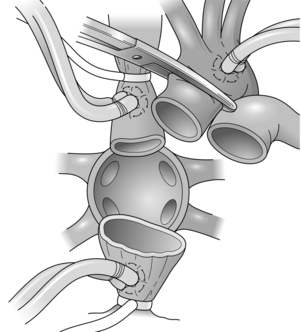
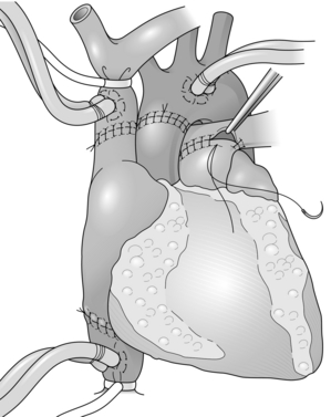
Heart transplantation (Figs 11.1 and 11.2)
The classical technique of orthotopic heart transplantation as described by Lower et al.32 and now modified to use the bicaval addition has remained the standard operation for 30 years.
Special situations
Heart transplantation for congenital heart disease
This group of patients presents special technical challenges due to unusual anatomy, previous operations and raised pulmonary vascular resistance. Heart transplantation can be undertaken to overcome most structural abnormalities.34
Heterotopic heart transplantation
1. if the pulmonary vascular resistance is high (pulmonary artery pressure > 60 mmHg) and cannot be manipulated by the use of nitric oxide;
The results of heterotopic transplantation have been generally inferior to orthotopic heart transplantation. However, this technique is occasionally considered.35
Perioperative management
< div class='tao-gold-member'>
Stay updated, free articles. Join our Telegram channel

Full access? Get Clinical Tree


