Introduction
Breast augmentation with primary fat grafting has gained increasing popularity in the past decade. It has several advantages over implant-based breast augmentation, including avoidance of foreign body implantation and related capsular contracture, which are appealing to people who want breast augmentation using autologous tissues. Initially, the safety and efficacy of fat grafting to the breasts had been questioned. However, a position paper from the American Society of Plastic Surgery Fat Grafting Task Force in 2009 concluded that fat grafting may be considered for breast augmentation and correction of defects associated with medical conditions and previous breast surgeries.
Several authors have also advocated the safety and efficacy of fat grafting to the breasts, and limited data on the radiologic impact of fat grafting to the breasts suggest that there is minimal interference with breast cancer screening. In addition, recent advancement in the concept and techniques of fat grafting has also added incentives to the popularity of this procedure. Proper patient selection is of utmost importance for a satisfactory outcome of breast augmentation with primary fat grafting.
In this chapter, the authors describe their preferred technique for breast augmentation with fat grafting, including patient selection, preoperative consultation, and several techniques to ensure an optimal outcome. In addition, a rationalized approach to fat necrosis after fat grafting for breast augmentation and an algorithm for management of fat necrosis are also introduced.
Indications and Contraindications
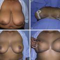
A good candidate for breast augmentation with fat grafting would be the one who has an abundance of body fat to be harvested from liposuction and sufficient skin pocket dimension or pliability for accommodation of adequate volume of graft. In addition, patients who desire a volume increase of 120–150 cc are good candidates because this procedure has limitations in possible volume increase after each session of fat grafting. There is no absolute contraindication for this procedure. However, patients who do not have sufficient fat to be harvested and those who have tight, small breast skin envelopes ( Fig. 6.1 ) and are reluctant to undergo either internal or external tissue expansion are not good candidates for this procedure. Box 6.1 summarizes the considerations for a good candidate for breast augmentation with primary fat grafting.
- •
A good abundance of fat in the body to be liposuctioned
- •
Good pliability and elasticity of skin in the breast
- •
Good volume of original breast tissue and pocket
Preoperative Evaluations and Special Considerations
Patients with a family history of breast cancer should be informed that the long-term safety of primary fat grafting to breasts has not been established in this group of patients and an extra effort in breast cancer surveillance may be needed after this procedure because of the possibility of calcification as a result of fat necrosis in the breast tissue. Fat necrosis may cause physical or psychological discomfort and potentially complicate breast cancer surveillance and should be communicated to the patient as a risk.
The volume of available fat is an important step for preoperative consultation and planning to ensure adequate volume to be harvested for single or multiple sessions of fat grafting depending on the patient’s expectation. Estimation of the volume of body fat is conveniently achieved by palm measurement in which one palm size is about 180–200 cm 2 depending on the surgeon’s palm size. The thickness of fat can be determined by a pinch test. The volume of fat to be collected is calculated by the area multiplied by the estimated thickness of fat that can be suctioned (volume = palm size 200 cm 2 × thickness of fat to be suctioned). In general, a total of 700–1000 cc of lipoaspirates (not including the infranatant) can be harvested from both thighs, which is enough for most patients.
The skin can be tested by finger stretching to see if it can be pulled off from the body with little resistance. Note especially the skin pliability in the lower pole, where it is most needed to be expanded for an anesthetic lower pole breast contour. If the skin envelope is tight and the original breast mound is small, then tissue expansion is necessary to overcome the skin tension to achieve the desired breast volume and shape. Tissue expansion can be either external or internal.
Patients who desire more than a two breast cup size increase should be informed of the necessity of more than one or multiple sessions of fat grafting. In patients who request breast augmentation by replacing previous breast implants with fat grafts, any breast deformity or asymmetry (e.g., capsular contracture) should be identified. If capsular contracture has developed, concomitant capsulotomy (Baker I–II) or capsulectomy (Baker III–IV) should be performed to achieve a smooth contour of the breasts ( Box 6.2 ).
- •
Assess the availability of fat volume in the body by palm test and skin pinch test.
- •
Check the pliability of the skin envelope of the breast by stretching the skin in the lower pole of the breast.
- •
Perform pre-expansion of the tight skin pocket of the breast using internal or external volume expansion before fat grafting.
- •
Subsequent grafting is necessary if more than a two breast cup size increase is expected.
- •
Capsulotomy or capsulectomy should be performed for capsule contracture in cases where fat grafting to breast is performed after implant removal.
Surgical Techniques
Relevant Surgical Anatomy
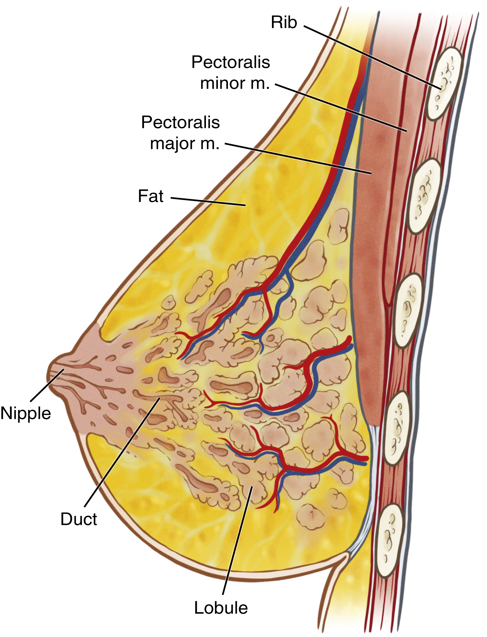
Injection of fat grafts to the breast requires an understanding of the relevant anatomy in this area for safety and efficacy of this operation. Fat grafts are injected in different depths of the breast mound: subcutaneous, breast tissue, under breast tissue, intra–pectoralis muscles, and subpectoral levels. Deeper to the subpectoral level lies the ribcage and intercostal muscles, and thus injection should not point down toward the thorax once the cannula is deep to subpectoral level because pneumothorax may occur once the cannula penetrates the pleura ( Fig. 6.2 ). The breasts are well-vascularized tissues and receive multiple blood supplies from several directions but mainly from perforating branches of the internal mammary artery. Therefore, gentle injection with a blunt-tipped cannula can minimize vessel injury and avoid major hematoma in the breast tissue.
Donor Site Selection
A variety of body areas that uniformly have abundant or excess fat are suitable as donor sites for harvest of fat grafts, such as the abdomen, flanks, buttocks, medial and lateral thighs, and knee. As a general rule, donor sites are selected that enhance body contour and are easily accessible in the supine position. Although the viability of adipocytes within the fat grafts from different donor sites may be considered equal, a higher concentration of adipose-derived stem cells (ADSCs) is found in the lower abdomen and inner thigh, which should therefore be chosen as the preferred donor sites for fat transplantation. ,
Anesthesia
The procedure is performed under general anesthesia or intravenous sedation. Intravenous sedation is routinely used in conjunction with regional or local anesthesia in the donor site of graft harvesting. The tumescent solution used for donor site analgesia or hemostasis should contain the lowest concentration of lidocaine because its high concentration may have a detrimental effect on adipocyte function and viability. In general, we often use 0.01%–0.02% of lidocaine in Ringer’s lactate if the fat grafting procedure is performed under general anesthesia. The tumescent solution also contains epinephrine with a concentration of 1:200,000. Epinephrine can precipitate vasoconstriction in the donor sites as well as the recipient sites, which may decrease blood loss, bruising, hematoma, and the possibility of intraarterial injection of the transplanted fat.
Fat Graft Harvesting
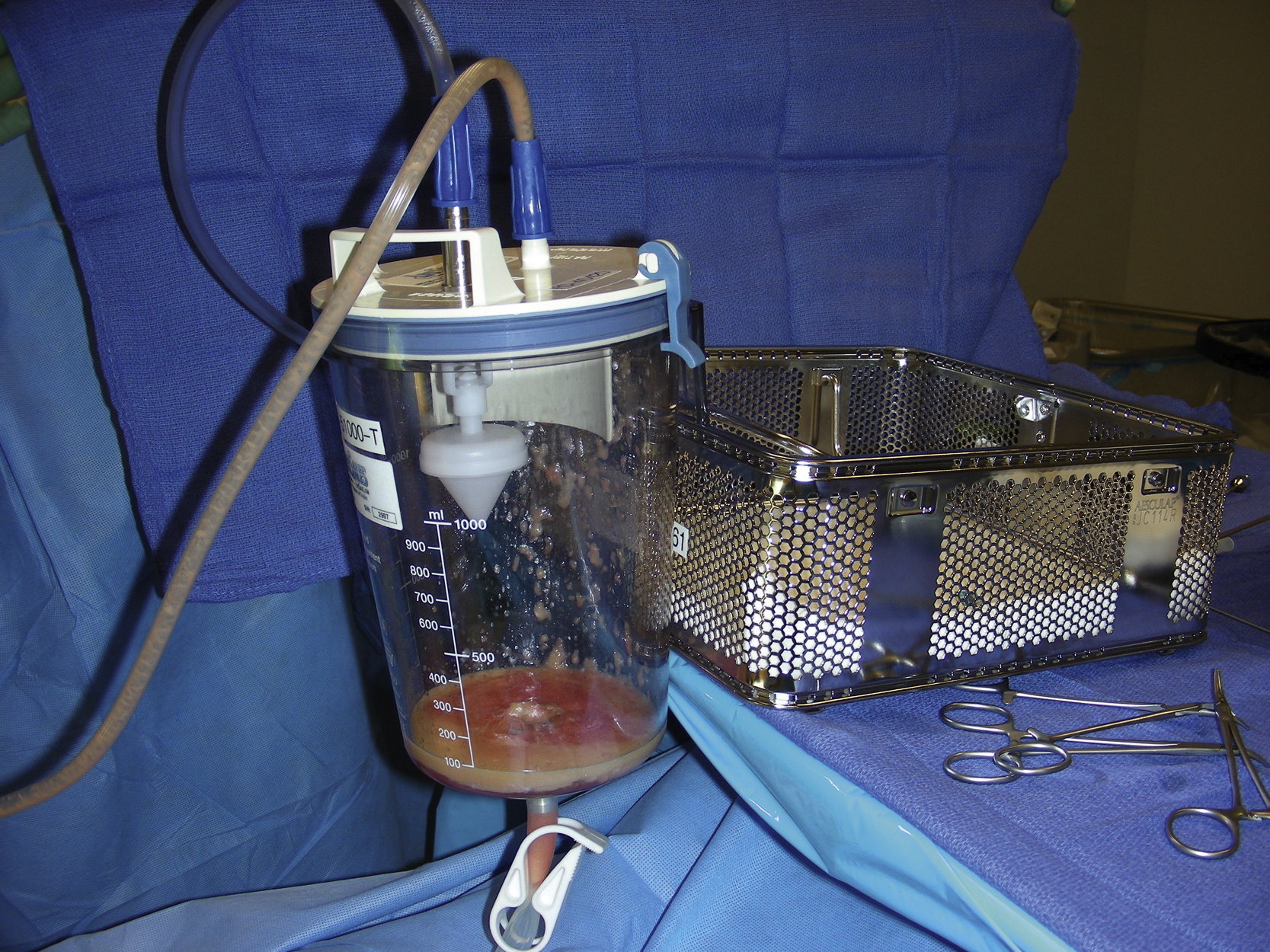
Placement of incisions can be done with a no. 11 blade in the locations where the future scar can be easily concealed. Fat grafts can be harvested through the same incision made for infiltration of anesthetic solution. The size of incision is about 3 mm. A tenotomy scissor is used to dilate the underlying subcutaneous tissue through the incision to allow insertion of the harvesting cannula with ease. The anesthetic solution is then infiltrated to the donor site 10–15 minutes before fat extraction, which makes harvesting of the fat graft easier and less traumatic. The ratio of aspirated fat to tumescent solution should be about 1:1 so that each pass of fat extraction can be more efficient. Vacuum-assisted liposuction with a machine is set to a pressure of –60 to –70 cm H 2 O. Lipoaspirates are collected into a 2-L canister that has a drainpipe with a lock attached to its bottom ( Fig. 6.3 ). The infranatant portion at the bottom of the canister is drained out through the pipe after lipoaspirate is allowed to settle for a while by gravity. The fat portion of the lipoaspirate is then collected easily through the pipeline into syringes of variable sizes of surgeons’ preferences for convenience of centrifugation.
Fat Graft Processing
To effectively remove the infiltrated solution and cell debris within the lipoaspirates and to obtain more concentrated fat grafts, centrifugation is our preferred method to process fat grafts. Recent studies have shown that proper centrifugation can concentrate not only adipocytes and ADSCs but also several angiogenic growth factors within the processed fat grafts.
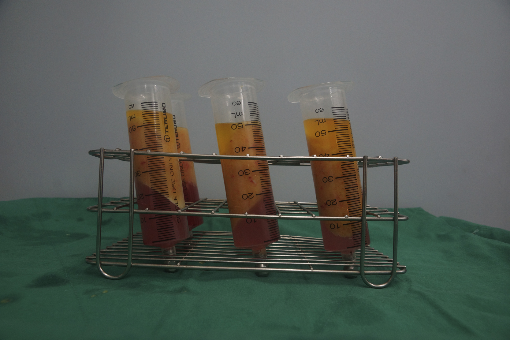
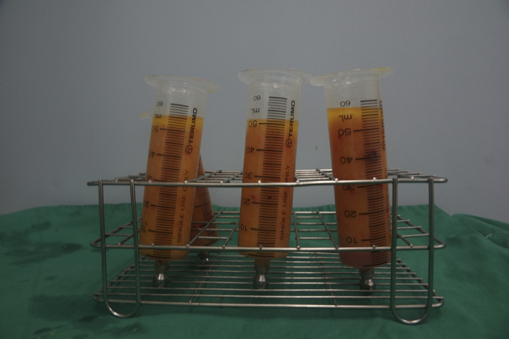
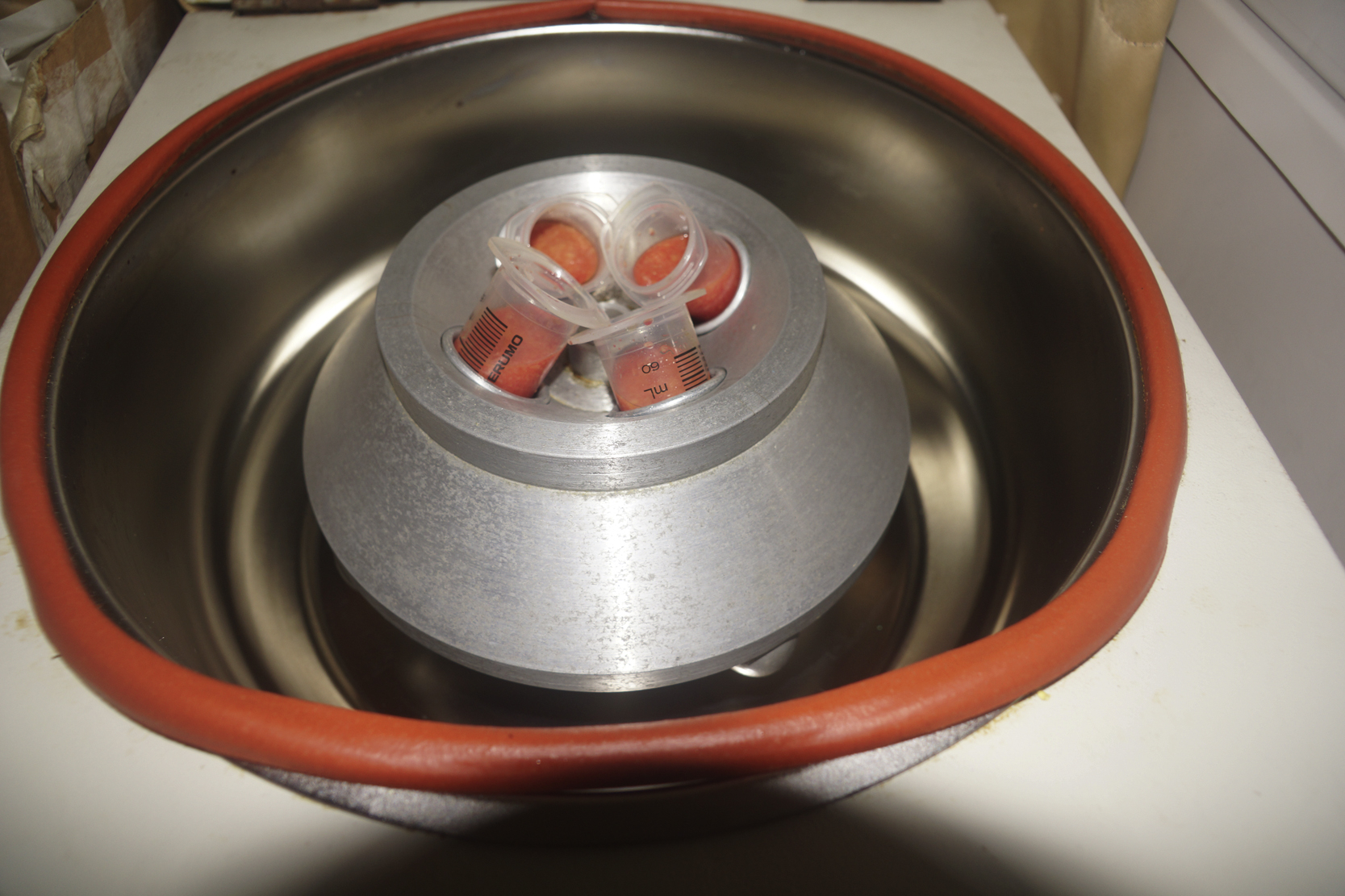
The authors choose a 50-cc Luer-Lok syringe for more efficient fat graft processing in mega-volume fat grafting. The fat portion of the lipoaspirates can be easily transferred to the 50-cc Luer-Lok syringe, which is snugly attached to the flexible hosepipe that drains out the lipoaspirates. The Luer-Lok aperture of the 50-cc syringe is then locked with a plug ( Fig. 6.4 ). After careful removal of the plunger, all lipoaspirate-filled 50-cc syringes are then centrifuged with 3000 rpm (about 1200 g ) for 3 minutes ( Fig. 6.5 ). A greater g force or longer duration of centrifugation may be harmful to adipocytes and is therefore not recommended.
In the authors’ experience, centrifuged lipoaspirate can be concentrated into 60% of its original volume. Therefore, about 700–1000 cc of lipoaspirate is needed at the completion of harvest because it can be concentrated into 400–600 cc fat for injection after centrifugation. The “no touch” method is preferred for fat graft processing because exposure of fat grafts to air and contamination can be avoided. After being centrifuged, lipoaspirates with the syringe are divided into three layers: the oil content in the upper layer, fatty tissue in the middle layer, and the fluid portion at the bottom ( Fig. 6.6 ). The oil can be decanted from the Luer-Lok syringe. The residual oil is wicked with a cotton strip or swab. The infranatant fluid at the bottom can be easily drained out once the plug at the Luer-Lok aperture is removed. The concentrated fat in the 50-cc syringe can then be transferred to a 10-cc syringe (our preferred size of syringe for fat injection in primary fat grafting to breasts) with an adaptor ( Figs. 6.7 and 6.8 ) . Attention should be given to the air bubbles inside the syringe because they could be removed and thus quantification of the volume injected can be recorded precisely.