Introduction
Breast reduction can be performed for either functional or cosmetic reasons. Optimal size, shape, and symmetry and minimal scarring, as four primary goals, should be applied to any type of breast reduction; for example, the breasts after reduction should be as the patient desired and in proportion to the patient’s body habitus. The shape after breast reduction should be cosmetically pleasing and hopefully long-lasting. Symmetry may be also important for most women after breast reduction.
Inverted-T inferior pedicle breast reduction was popularized in the 1970s in the United States. It is still the most commonly performed breast reduction procedure in the United States. The procedure itself can be suitable for almost all patients and in various breast sizes and shapes. Its design and surgical technique are reasonably consistent, and it can be performed in a standardized fashion. It is considered the most versatile but safe technique for breast reduction, with lower rates of complication or revision, , although prominent scarring or “bottoming-out” can be a concern over the long term.
This chapter describes the author’s preferred technique for inverted-T inferior pedicle breast reduction. Several technical refinements of the surgical technique are described in detail. In addition, pearls to achieve an optimal outcome and management of complications after inferior pedicle breast reduction are discussed.
Indications and Contraindications
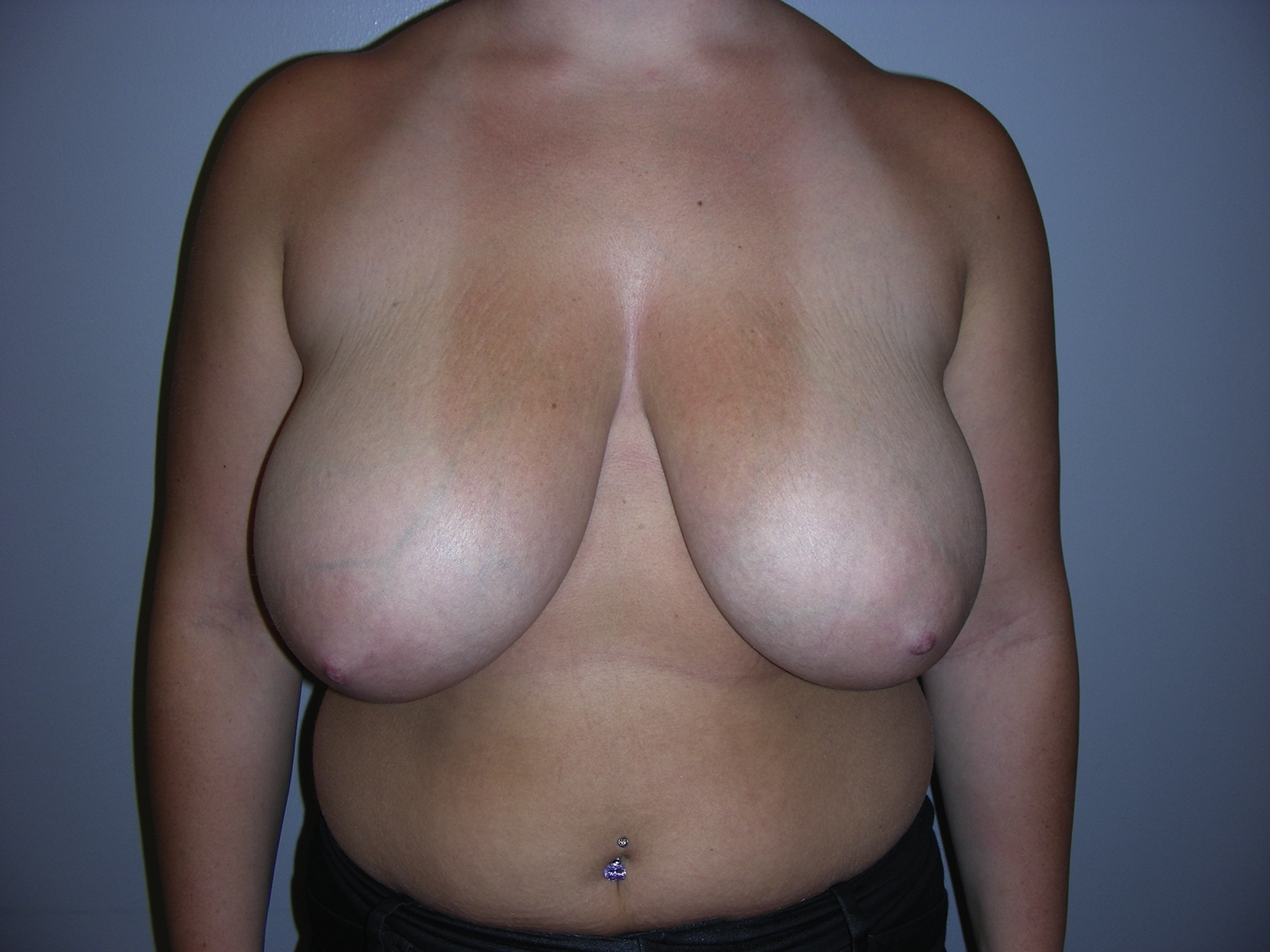
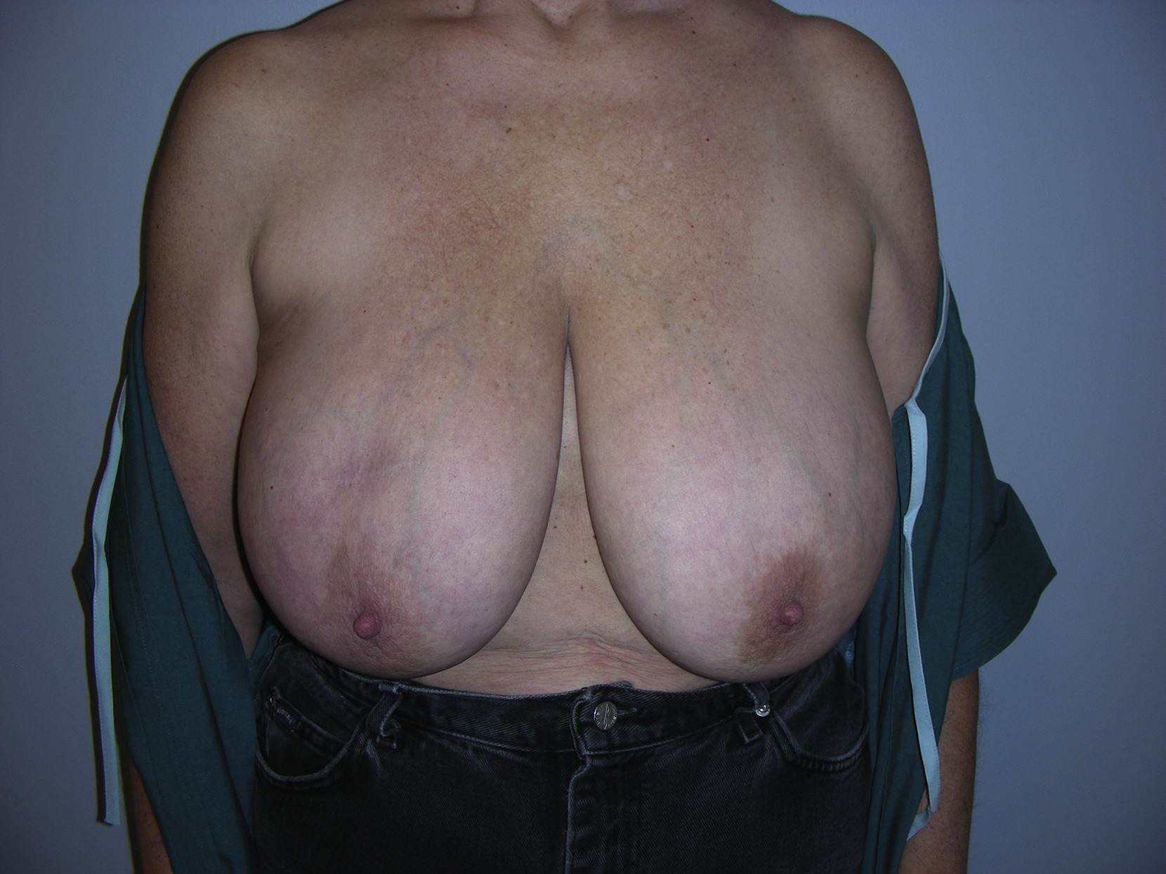
It is a common thought that classic inferior pedicle breast reduction is indicated for almost all patients regardless of breast size and shape ( Fig. 19.1 ). For patients who are relatively older and have an elongated breast shape because of poor breast skin condition, the inverted-T inferior pedicle breast reduction can be selected for more predictable results ( Fig. 19.2 ). The overall amount of breast tissue reduction may not be critical, although the average weight of this type of breast reduction is usually less than 1000 g from each breast. However, the distance from the suprasternal notch to the nipple should be less than 15 cm so that adequate blood supply to the nipple can be ensured based on the inferiorly based pedicle. If the distance is more than 15 cm, a free nipple graft procedure should be considered based on common standard practice. In general, the inverted-T pattern will remove excess breast skin from both vertical and horizontal orientations. Younger women with good breast skin condition (no stretch marks) may have a better long-term outcome, although the inverted-T inferior pedicle breast reduction has been criticized as resulting in a bottoming-out breast shape and an unsightly scar.
Preoperative Evaluation and Special Considerations
Unlike the medial pedicle breast reduction technique, the classic inverted-T inferior pedicle breast reduction has fewer special considerations and intraoperative adjustments. Each step of the procedure can be performed in a standardized fashion based on the preoperative and intraoperative markings ( Fig. 19.3 ). However, several important points should be considered to achieve an optimal outcome after the inferior pedicle breast reduction. The new nipple position should be placed 1 cm lower than the level of the inframammary fold (IMF) to avoid a possible high-riding nipple. The inferior pedicle should be made sufficiently thick and may include the perforators from the central part of the breast to ensure robust blood supply to the nipple. The plication of the inferior pedicle can ensure upper pole fullness and easy in-set of the nipple–areola complex. The lateral horizontal incision should not be extended beyond the anterior axillary line for most cases.
The distance between the nipple and the IMF should be controlled to 5–6 cm for the classic inverted-T inferior pedicle breast reduction so future bottoming-out may be avoided. However, this also depends on patient breast skin quality.
In management of the inverted-T closure in the lower pole of the breast, it is important to ensure primary healing because of the tension in this area after the closure. The surgeon should pay attention to this important issue and develop a strategy to reduce tension on the closure ( Box 19.1 ).
- •
Procedure can be performed for all patients regardless of breast size and shape.
- •
It is a good choice for patients with elongated shape (severe ptosis) of the breast.
- •
Various amounts of breast reduction can be accommodated up to 1000 g for each breast.
- •
The distance from the suprasternal notch to nipple should be less than 15 cm.
- •
Proper intraoperative management of the pedicle size, shape, and length is important.
- •
Proper design of skin pattern and management of the inverted-T closure is important.
- •
Prominent scar and bottoming-out of the breast can be a problem.
Surgical Techniques
Relevant Surgical Anatomy
The relevant anatomy of the breast has been described in Chapter 18 . Once again, the blood supply to the breast in general comes in from several directions. However, the main blood supply to the breast is based on the medial branches of the internal mammary vessels. The inferior pedicle receives its blood supply from the perforators through the pectoral muscle that come from the internal mammary vessels at the fourth intercostal space and may be accompanied by venae comitantes. There, perforators enter the breast just medial to the breast meridian approximately 4–6 cm above the IMF and provide adequate blood supply to the pedicle and nipple–areolar complex as long as adequate width and thickness of the pedicle can be maintained ( Fig. 19.4 ). Again, the nipple is primarily innervated by the medial and lateral branches of the fourth intercostal nerve. However, the third and fifth intercostal nerves may contribute as well.
Preoperative Markings
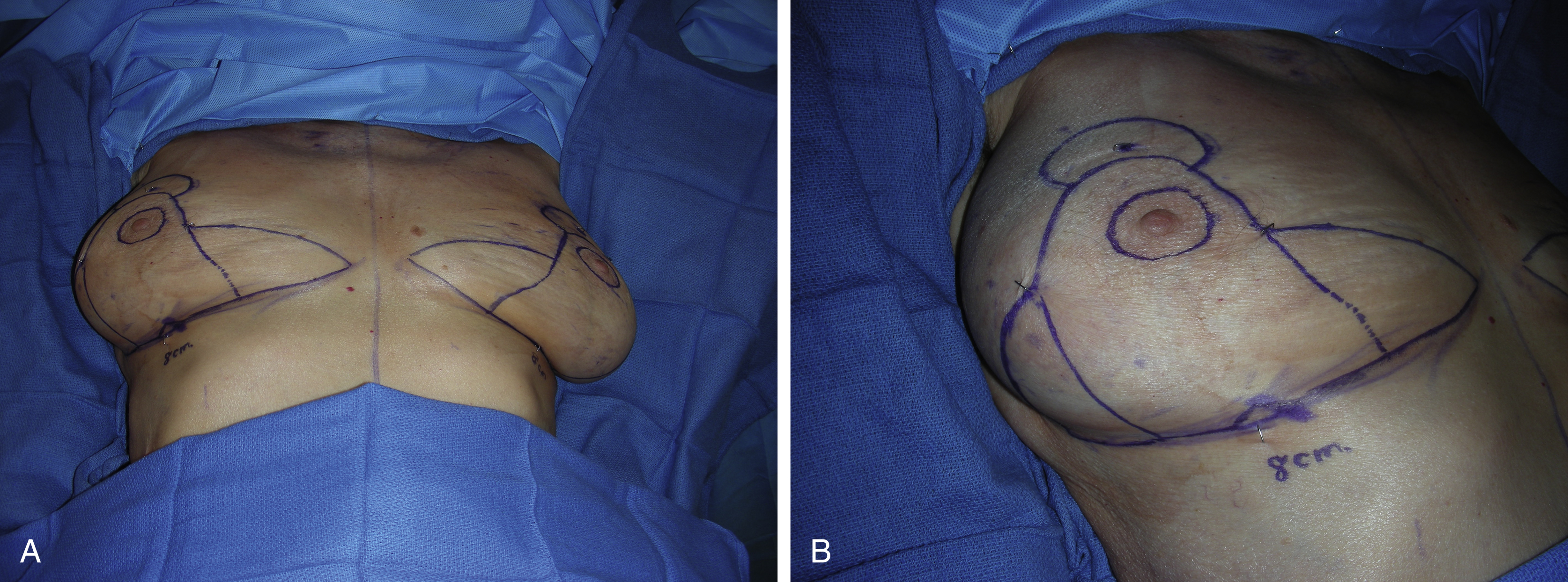
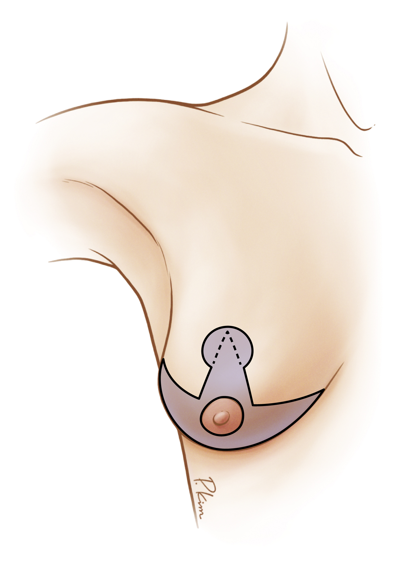
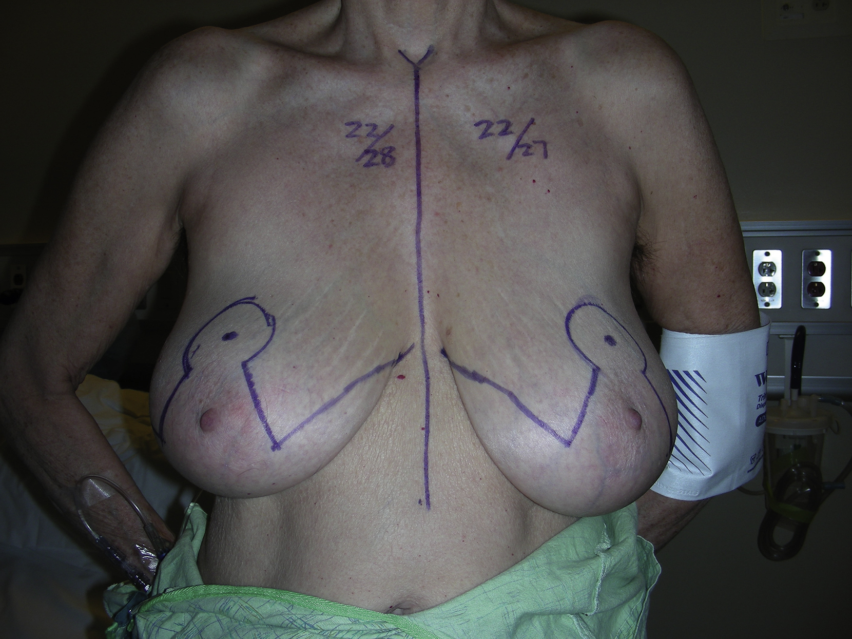
While the patient is in the upright position, the new nipple position is marked first. For the classic inverted-T inferior pedicle breast reduction, the new nipple position should be set 1 cm below the level of the IMF so that a high-riding nipple position can be avoided. This author’s preference is to use a Wise pattern template for all initial marking in a standard fashion. The distance for each vertical length should be less than 6 cm (usually 5 cm), and the inverted-T closure could be approximated without tension. For horizontal marking along the IMF, the medial extent of the incision should be away from the midline of the anterior chest and should not be visible while the patient is standing. The lateral extent of the incision should not go beyond the anterior axillary line of the patient and should not be visible. Attention should be given to symmetric placement of the new nipple position in each breast based on gross inspection and measurements ( Fig. 19.5 ).
Intraoperative Markings
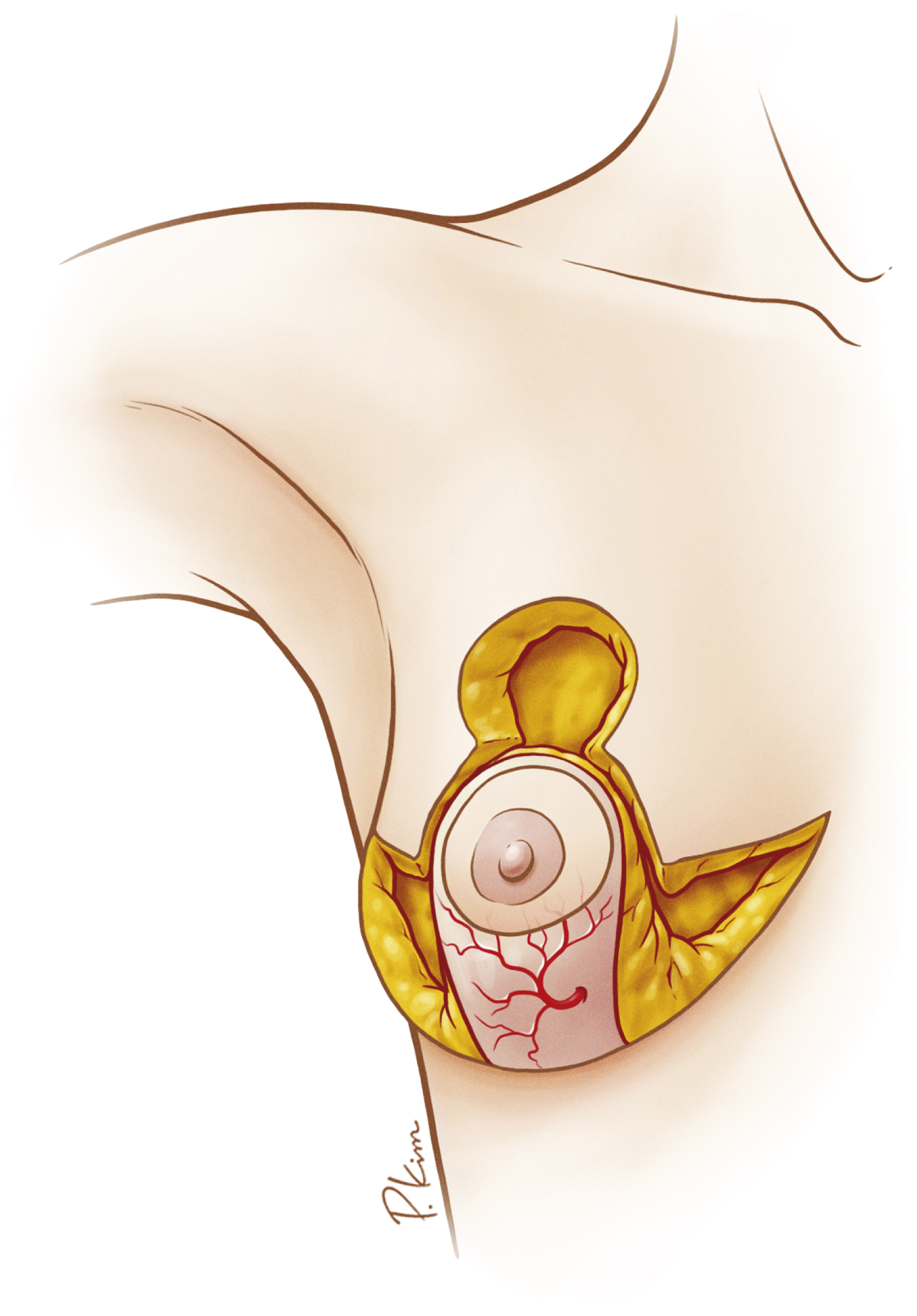
While the patient is in the supine position, commonly under general anesthesia, the nipple–areola complex is marked with either a 38- or 42-mm cookie cutter based on the location of the new nipple position. Once the midline of each breast is determined along the IMF the inferior pedicle can then be designed, with the pedicle width from 7–9 cm depending on the breast size the surgeon wants to achieve after breast reduction. It is important to leave at least a 1-cm-wide section of breast tissue from the proposed upper border of the nipple–areolar complex to avoid cutting into it ( Fig. 19.6A ). After completion of the pedicle marking, a small triangle is marked in the middle of the horizontal inferior pedicle to serve as a skin bridge to potentially reduce tension on the inverted-T closure (see Fig. 19.6B ). Finally, the marked medial and lateral vertical limbs in the breast should be tested for easy approximation in the proposed inverted-T closure without tension.