Bites and Stings


Bites and Stings


Tracy Lynn Donahue


SPIDERS


I. BACKGROUND

Spider bites are common, but most species cause minimal or no harm to humans. Approximately 14% of deaths from venomous animals result from spider bites. The species that most commonly cause severe reactions in humans in the United States are the black widow spider (Latrodectus mactans) and the brown recluse spider (Loxosceles reclusa).

The black widow spider is found throughout the United States and commonly lives in wood piles. The species is identified by a distinct red hourglass on its abdomen. It injects venom containing the neurotoxin α-latrotoxin into its prey. α-Latrotoxin destabilizes nerve cell membranes by opening ionic channels, causing irreversible acetylcholine release.

The brown recluse spider is endemic to the Midwest United States. Geographical distribution may be expanding with climate change, although many reports of bites are likely due to spider misidentification or misdiagnosis. The brown recluse spider has long legs compared with its body. It is recognized by its yellow-brown color and violin string marking on its back. The brown recluse spider injects sphingomyelinase D which triggers platelet aggregation, endothelial hyperpermeability, hemolysis, and neutrophil-induced skin necrosis. It typically only bites when threatened.


II. CLINICAL PRESENTATION

Black widow bites are sensed as a pinprick, followed by a dull aching pain. After about 30 minutes, the skin surrounding the bite may feel numb. The skin appears erythematous and mildly edematous with local piloerection. In addition, there may be local perspiration and lymphangitis. Peaking between 1 and 8 hours after the bite, a systemic anticholinergic response may last up to 2 days (Table 6-1). A characteristic facial swelling, known as Latrodectus facies, can also be seen. Death occurs in less than 1% of all black widow bites.

Brown recluse bites cause local reactions that range from a mild local burning sensation to severe cutaneous necrosis. Severe reactions are seen in less than 10% of bites. Severe reactions generally begin as localized edema with an erythematous halo, resembling a “bulls-eye.” Over the next 12 hours, bullae form centrally. The plaque forms an irregularly shaped blue-black eschar, surrounded by pallor and a larger area of erythema, forming the characteristic “red, white, and blue sign.” Less serious lesions heal over weeks while more severe bites heal over months and may leave a residual scar. Associated systemic symptoms are listed in Table 6-1. Death may result from acute renal failure or intravascular hemolysis, usually 2 to 3 days following the bite.


TABLE 6-1 Systemic Symptoms to Spider Bites


Black Widow Spider (toxin = α-latrotoxin)


Miosis


Lacrimation


Sialorrhea


Hyperhidrosis


Bronchospasm


Bradycardia


Abdominal cramping and diarrhea


Increased urination


Skeletal muscle spasm and rigidity


Brown Recluse Spider (toxin = sphingomyelinase D)


Fever


Morbilliform rash


Arthralgias


Nausea and vomiting


Respiratory failure (pulmonary edema)


Anemia due to intravascular hemolysis


Disseminated intravascular coagulation


Renal failure


Rhabdomyolysis


III. WORKUP

Diagnosis of a spider bite can be difficult clinically due to a broad differential diagnosis (Table 6-2). Ideally the offending spider is captured for identification. Wounds can be cultured to evaluate for superinfection. Skin biopsy may reveal neutrophilic perivasculitis, hemorrhage, edema, necrosis, and a prominent eosinophilic and neutrophilic infiltrate which may involve the subcutaneous fat. Workup for systemic involvement should include a sedimentation rate, complete blood count, fibrinogen, coagulation studies, urinalysis, and renal function studies.


SNAKES


I. BACKGROUND

Between 4,000 and 6,000 venomous snakebites occur annually in the United States. The two families of snakes of medical importance in the United States include the Viperidae and Elapidae families.

The Viperidae family, or pit viper snakes, include rattlesnakes, cottonmouth (water moccasin), and copperhead varieties. They are recognized by their triangular head, elliptical “cat’s eye” pupils, single row of ventral scales,
and depressed heat-sensing facial pit. Eighty percent of bites result in injection of venom. Pit viper venom has hemorrhagic and coagulopathic properties and may contain presynaptic neurotoxins. Bites from the diamondback rattlesnake account for 95% of Viperidae fatalities.

The Elapidae family includes the coral snake whose venom causes fibrinolysis, anaphylaxis (via activation of the complement cascade), and neurotoxicity. They are recognized by their round eyes, and red, yellow, or white bands. In the United States, they mimic the nonvenomous King snake which has red and black bands. The distinction can be remembered by “Red on yellow, kill a fellow; red on black, venom lacks.”


II. CLINICAL PRESENTATION

Pit viper bites result in immediate local pain and visible fang puncta. Initial weakness, swelling, paresthesias, nausea, and vomiting ensue. Local reactions due to direct tissue injury and victim cytokine responses include ecchymoses, tenderness, bullae formation in flexure creases, and myonecrosis. Local reactions spread via lymphatics. Less common complications may include compartment syndrome and wound infection. Venom may rarely induce urticaria, angioedema, and anaphylactic reactions. Hematologic venom effects include fibrinogen degradation and destruction, manifesting as bleeding at the bite site, gingiva, or nasal mucosa. More severe presentations include gastrointestinal or intracranial hemorrhage. Systemic venom effects may cause direct cardiovascular toxicity, respiratory arrest, disseminated intravascular coagulation, acute renal failure, neurotoxicity, and rhabdomyolysis.

Coral snake bites are typically painless without local edema or necrosis. The neurotoxic effects cause limb weakness or numbness in the bitten extremity. Within hours, systemic symptoms including tremors, muscle fasciculations, sialorrhea, and bulbar paralysis (causing dysphagia, dyspnea, or total flaccid paralysis) ensue. Paralysis of the diaphragm leads to respiratory paralysis and death.


III. WORKUP

Snake envenomation victims should be assessed and observed with serial examinations in facilities prepared to handle acute hematologic, neurologic, respiratory, and cardiovascular emergencies. Lab work should include type and cross, complete blood count, prothromin time, partial thromboplastin time, fibrinogen levels, and fibrin split products.


INSECTS


I. BACKGROUND

The most serious reactions to insects are caused by acquired hypersensitivity. Over 80% of deaths from insect bites and stings result from anaphylactic reactions and occur within 1 hour of the event. Anaphylaxis occurs in 3% of adults and 1% of children after an insect sting. Many patients who develop generalized reactions to insect bites or stings have no history of previous systemic of local reactions to the insect. Patients with a large local reaction incur only a 5% to 10% chance of developing a systemic reaction.


TABLE 6-4 Treatments to Avoid Following Pit Viper Snakebites


Treatment to Avoid


Reasons for Avoidance


Wound incision and suction


Does not remove significant amounts of venom


Worsens local tissue injury


Ice and cryotherapy


Associated with severe iatrogenic tissue injury


Tourniquet and pressure immobilization


May improve or worsen outcomes


Electrical current


Ineffective


Causes significant tissue injury


Prophylactic fasciotomy


Antivenom alone may reduce compartment pressures


Nonsteroidal anti-inflammatory drugs


Theoretical harm from platelet dysfunction in a thrombocytopenic patient


Prophylactic antibiotics


Leads to unnecessarily prolonged hospital stays


Corticosteroids


No evidence of improved outcomes


The most venomous insects belong to the order Hymenoptera, which includes bees, wasps, and ants. Hymenoptera venom contains serotonin, kinins, acetylcholine, lecithinase, hyaluronidase, phospholipase, and melittin. Exposure to venom in allergic individuals causes histamine release from leukocytes.

The order Diptera includes mosquitoes and flies. Mosquitoes inject salivary fluid into the victim, causing an immediate allergic reaction. Flies pierce through the skin, directly inserting saliva that causes both allergic and toxic reactions.

The order Hemiptera includes the bed bug (Cimex lenticularis) and the Reduviid bug. Historically, bed bugs were found in poor living conditions and unkempt hotels. In recent years, infestations have increased at a rapid rate and now include suburban and higher income populations as well. Bed bugs hidden in mattresses and upholstery come out at night to feed. The Reduviid bug of Mexico, Central America, and South America lives in cracks of mud hut walls and spreads disease-depositing stool on the skin as it bites.

The Meloidae and Staphylinidae families of the order Coleoptera include blister beetles. Blister beetle species of varying colors and patterns are found worldwide, with approximately 300 species residing in the United States. When blister beetles are pressed or rubbed, pederin- or cantharidin-containing hemolymph blisters the skin. Cantharidin is a toxin of medical interest, and it is used for treatment of warts and molluscum in dermatology.


II. CLINICAL PRESENTATION

A. Hymenoptera. Hymenoptera stings usually result in instantaneous pain followed by a localized wheal and flare reaction with associated pruritus. Edema is variable (Fig. 6-1). Multiple bee, wasp, yellow jacket, or hornet
stings can result in systemic reactions, including vomiting, diarrhea, generalized edema, dyspnea, hypotension, and collapse. Life-threatening allergic sting reactions are characterized by the usual manifestations of anaphylaxis: urticaria, laryngeal edema, bronchospasm, abdominal cramps, and shock. Harvester and fire ants are known for the intense burning and pain associated with their stings. Localized necrosis secondary to phospholipase and hyaluronidase in the venom may be present at the sting site.


Figure 6-1. Angioedema due to a bee sting. This patient developed an immediate hypersensitivity reaction after being bitten on the lip. (From Goodheart HP. Goodheart’s Photoguide of Common Skin Disorders. 2nd ed. Philadelphia, PA: Lippincott Williams & Wilkins; 2003.)

B. Diptera

1. Mosquitoes. Mosquito bites result in immediate allergic reaction to injected salivary fluids. Several hours later, a pruritic papule develops. Mosquitoes can also serve as the host for diseases, including Eastern and Western Equine Encephalitis, West Nile Virus, filariasis, malaria, and yellow fever.

2. Flies. Flies bites cause immediate pruritic wheals followed by itchy, red papules. The black fly is renowned for inducing extremely painful and long-lasting reactions. Flies associated with disease are listed in Table 6-5.

C. Siphonaptera (Fleas). Bites usually appear as grouped urticaria papules, often with a central punctum. They are commonly located on the distal extremities. Fleas are vectors for the bubonic plague, brucellosis, melioidosis, erysipeloid, and endemic typhus.

D. Hemiptera (True Bugs). Bed bugs leave linearly arranged pruritic papules in groups of two or three on the face and extremities, known as the “breakfast, lunch, and dinner” sign. The Reduviid bug acts as a vector for Trypanosoma cruzi, causing American trypanosomiasis or Chagas disease. Known as the “kissing bug,” the Reduviid often bites the victim’s face. Local reactions include a unilateral periorbital conjunctivitis and periorbital edema, known as the “Roma-a sign,” and regional lymphadenopathy
known as the “Chagoma.” Systemic reactions include myocardial damage, megacolon, and megaesophagus.
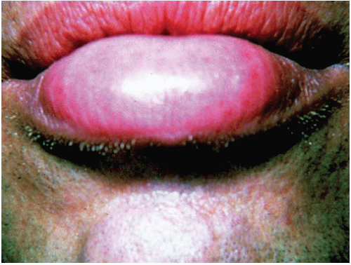


TABLE 6-5 Fly-Associated Disease


Only gold members can continue reading. Log In or Register to continue

Stay updated, free articles. Join our Telegram channel

Jun 10, 2016 | Posted by in Dermatology | Comments Off on Bites and Stings

Full access? Get Clinical Tree

Get Clinical Tree app for offline access

Fly


Geography


Disease


Clinical Features


Botfly


(Dermatobia hominis)


Mexico Central America South America