15 Basic Local Perforator Flaps of the Lower Extremity
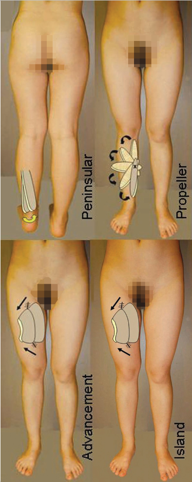
Fig. 15.1 The fundamental subtypes of local perforator flaps.
Summary
Some consider a perforator flap to be a variant of a fasciocutaneous flap that has a specific circulation pattern arising from a known vessel that has perforated the deep fascia. The lower extremity has numerous such perforators that arise in predictable regions or “hot spots,” but are individually highly variable. Each supplies a perforasome that can be the basis of a local flap. Local flaps have had increasing popularity as a means to avoid the detriments of free flaps. Yet it must be appreciated that even the harvest of local perforator flaps still requires microsurgical skills, although there need not be a microvascular anastomoses. There are four basic subtypes of local perforator flaps as defined using the traditional nomenclature. These are the peninsular, V-Y or keystone advancement, propeller, or true island flaps that have a lengthy vascular pedicle. Important variants of this theme include the plantar flaps, valuable for solving difficult glabrous sole challenges, and the distal-based sural flap that can similarly be useful without the need for microvascular surgery.
Keywords: perforator flap, peninsular flap, keystone flap, propeller flap, island-pedicled flap, medialis pedis, distal-based sural flap
15.1 Introduction (▶ Fig. 15.1)
Some might say “back to the future.” Old-fashioned random flaps survived “randomly” on a subdermal vascular plexus of unknown origin.1 They could be distinguished from each other only according to their means of transposition (e.g., advancement or rotation)1 or their geometrical configuration (e.g., as a tubed flap).2 Strict length-to-width ratios were enforced to ensure reliability, and varied depending on the body region. Methods to delay random flaps for their enlargement or greater reach required multiple time-consuming stages.3 Milton4 then abruptly destroyed these concepts demonstrating that the source of circulation was the most important factor for flap survival.
McGregor and Morgan5 proved Milton right, pointing out that flaps with an intrinsic arteriovenous network would be viable to greater lengths as an axial flap. Even then as shown in their statement regarding the deltopectoral axial flap, they were aware that its network was nourished by “perforating branches of the internal mammary system.”6 Bigger skin flaps without a delay could also be immediately obtained by inclusion of the underlying muscle as a musculocutaneous flap.7 Then Pontén8 reintroduced the fasciocutaneous flap that had neither muscle nor an axial pedicle, yet achieved similar results. Although Pontén never specified the source of circulation to his superflaps,8 Cormack and Lamberty9 in their classification schema for fasciocutaneous flaps nicknamed them as being random flaps, but they understood well that their circulation relied on fascial feeders with multiple inputs to a fascial plexus. Fasciocutaneous flaps as such became valuable throughout the lower extremity,10,11 and that of Donski and Fogdestam12 probably was the precursor to the distal-based sural flap.
But Cormack and Lamberty also knew their other fasciocutaneous flap subtypes differed, since they were nourished by discrete septocutaneous perforators.9 Song et al13 published the anterolateral thigh flap also as a septocutaneous perforator perfused flap, although today more often usually this has been found to be via a musculocutaneous perforator. The latter would qualify as a “true” perforator flap,14 and this genre was soon widely used primarily as a free flap.15 Wei and Mardini,16,17 in the search for the perfect donor site, believed that only an adequate perforator needs to be present there, about which could be designed the so-called free-style free flaps. Yet why not use these same perforators in this “free-style” fashion to also be the pedicle to a local flap in the lower extremity in lieu of more complex free flaps?18,19 So began a gradual transition in that direction with a comparison of the risks and benefits in using local versus free perforator flaps just in the lower extremity.20,21 No significant difference was found, as long as it was appreciated that from a technical standpoint, a local perforator flap had to be carefully harvested as a microsurgical yet nonmicrovascular tissue transfer.22 To simplify the technique for the construction and nomenclature for these lower limb local perforator flaps, Lu et al23 classified them into four subtypes leaning on traditional terminology. Their simplest form was the peninsular flap, which has limited rotation.24 The V-Y25,26 or keystone flaps27 are those most practical for advancement. The propeller flap version that Hyakusoku et al introduced, based on a random subcutaneous pedicle,28 in the lower limb is more useful if the hub instead is a perforator.24,29 Finally, a “true” island flap has a pedicle that extends beyond the perforator to allow astounding reach.30,31
There can be no question, and for whatever reasons, a paradigm shift has occurred with the preferred selection of local perforator flaps whenever feasible for coverage problems throughout the lower extremity.32 This is the rationale here for further emphasis on the fundamental subtypes of local perforator flaps pertinent for use in the lower limb, as well as an additional bonus about the role of plantar flaps and the distal-based sural flap for foot and ankle soft-tissue closure. Anatomists have found that the subdermal vascular plexus that nourished the random flaps of past eras in truth intimately relied on deep fascial perforators. No longer just random flaps, these now are actually perforator flaps. The history of flaps has come full circle.
References
[2] Webster JP. The early history of the tubed pedicle flap. Surg Clin North Am. 1959; 39(2):261–275
[3] Bowen J, Meares A. Delayed local leg flaps. Br J Plast Surg. 1974; 27(2):167–170
[5] McGregor IA, Morgan G. Axial and random pattern flaps. Br J Plast Surg. 1973; 26(3):202–213
[6] McGregor IA, Jackson IT. The groin flap. Br J Plast Surg. 1972; 25(1):3–16
[11] Hallock GG. Local knee random fasciocutaneous flaps. Ann Plast Surg. 1989; 23(4):289–296
[17] Wei FC, Mardini S. Free-style free flaps. Plast Reconstr Surg. 2004; 114(4):910–916
15.2 Chapter 15A: The Island Perforator Flap
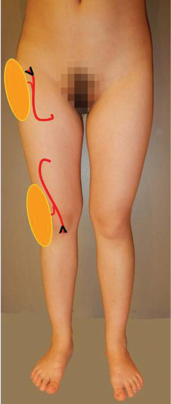
Fig. 15.2 Proximal and distal-based versions of an island flap.
15.2.1 Introduction to the Island Perforator Flap
Just what is a “true” island perforator flap (▶ Fig. 15.2) is a highly controversial idea. Theodore Dunham1 (1892) is credited with using the term clinically for the first time, and he defined an island flap as one having an axial pedicle that is skeletonized and then tunneled under a cutaneous bridge to allow coverage of a nonadjacent defect. Today the term has become less specific, often applied to any flap whose peripheral boundaries are separated in toto from its donor site,2,3 remaining attached only by whatever is its source of vascularization. For example, a V-Y or keystone “island” advancement flap remains attached only by a broad base to the underlying deep fascia.4 A propeller perforator flap is connected only at its hub, that typically being a single perforator that at most has been dissected back to its source vessel to form what is often called an island flap. Rather than an “island” flap, which is somewhat of a misnomer, a better term for the latter two examples might be “islanded” flaps.
Yet Dunham’s1 definition still has merit for describing this subtype of lower limb local perforator flaps. What is a “true” island perforator flap must be approached somewhat differently in order to obtain its somewhat unique attributes. Not only must all skin margins be disconnected at the donor site, but also the axial pedicle providing the flap circulation must be extended beyond just the requisite diminutive perforator to include a major branch if not the source vessel itself. Early examples obeying Dunham’s1 definition in the lower leg were fasciocutaneous flaps that indeed were sustained by perforators from the anterior tibial,5 posterior tibial,6 or peroneal7,8 vessels, but these source vessels themselves were also elevated with the flap over considerable distances. For foot and ankle defects, usually these major vessels would be divided just proximal to the connected perforator-based flap, then the source vessel pedicle dissected distal to the desired point of rotation that allowed appropriate flap insetting. Reverse flow was essential for flap survival, and the source vessel itself was sacrificed irrespective of any risks of long-term sequela. The omnipresence of peripheral vascular disease would make this option unacceptable today, and fortunately many of the other perforator local flap subtypes can now provide the same benefit.
Nevertheless, from a practical standpoint, several possibilities for “true” island flaps still exist in the lower extremity. Zheng et al9 have described use of a constant perforator of the descending genicular artery (DCA) to sustain a distal anteromedial thigh flap as a propeller flap, but this could also be an island flap for proximal thigh transposition if based instead on the DCA itself. Agko and Chen (Chapter 15E: Medialis Pedis and Other Plantar Flaps) have discussed several local plantar flaps that can similarly be extended on the medial plantar artery as island flaps to allow reach to many sites about the foot and ankle. However, two more pragmatic options in the lower limb are island anterolateral thigh (ALT) and medial sural artery perforator (MSAP) flaps. As is well known, the proximal-pedicled ALT island flap can reach superiorly to the lower abdomen, laterally the greater trochanter, and medially the perineum (▶ Fig. 15.3).10,11 Both a distal-pedicled ALT island flap (▶ Fig. 15.3)12 and a proximal-pedicled MSAP island flap13 (▶ Fig. 15.4) can cover the patella or the proximal lower leg. Because these “true” island flaps rely on inclusion of their source vessel, the descending branch of the lateral circumflex femoral (LCF) or medial sural artery, respectively, reach of these flaps is limited only by the potential length of that pedicle.
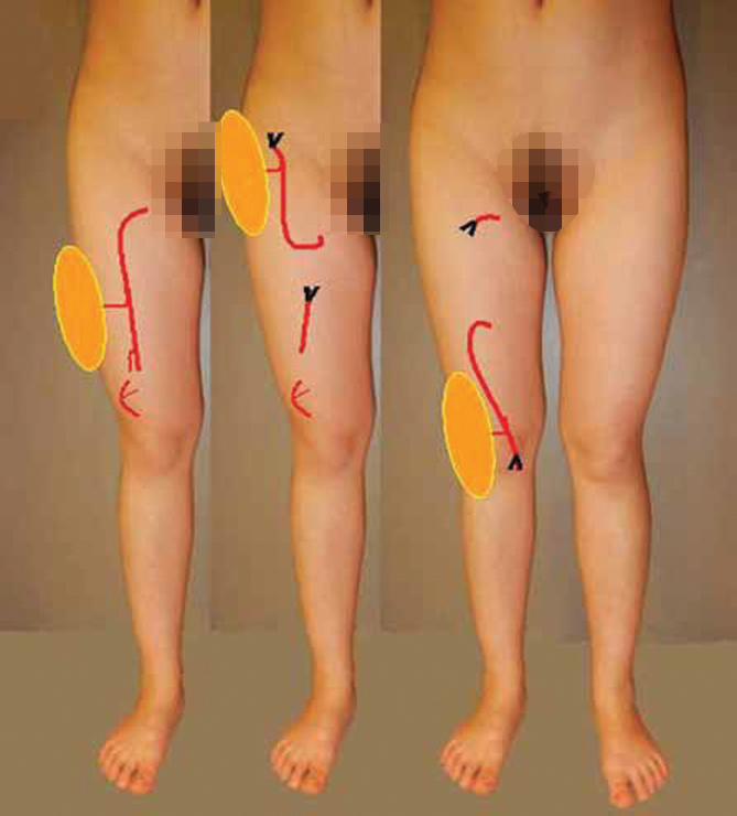
Fig. 15.3 This model of the anterolateral thigh (ALT) flap, based on a perforator of the descending branch of the lateral circumflex femoral, shows that a “true” island flap can have a long proximal pedicle for orthograde perfusion, or instead be based distally to rely on reverse flow. In either scenario, significantly greater reach will be possible than with any other pedicled perforator flap subtype.
15.2.2 Attributes and Detriments
Attributes
• Local flap.
• Normal contour and appearance.
• Long vascular leash with enhanced reach.
• Knee or hip coverage depending on flap selection.
• Function preservation.
• No microsurgery.
Detriments
• Extensive pedicle dissection.
• Sacrifice of source vessel.
• Risk of venous congestion with any distal-based variant.
15.2.3 Anatomical Considerations
To avoid redundancy, the basic anatomy of the ALT and MSAP flaps is no different than as catalogued in Chapter 16. As a local island flap, however, the difference will be the need to extend the source vessel pedicle while always being retained in situ at the point of flap rotation. For example, the pedicle of the MSAP flap can be lengthened to improve reach by dissection back to its origin from the popliteal vessels. The ALT flap pedicle similarly can be freed up back to the LCF vessels. Once this maneuver has been accomplished, passing the island ALT flap under the rectus femoris muscle will further increase reach to the midline or beyond.14 If the ALT flap is used as a distal-based flap, maintenance of communications of the descending branch of the LCF system, which presumably also services the flap perforator, with collaterals from the lateral superior geniculate vessels for reverse flow will be essential.12
15.2.4 Anatomical Variations and Potential Pitfalls
Sural Artery Flaps
Almost always a single major musculocutaneous perforator will be found exiting the medial head of the gastrocnemius muscle.15 The more distal the perforator, the longer the potential vascular pedicle to allow an MSAP island flap to reach the patella or the proximal tibia. A musculocutaneous perforator of the lateral head is often absent, so another alternative must always be anticipated.15
Distal-Based Anterolateral Thigh Flap
The source vessel of the perforator to the ALT flap may not be the descending branch of the LCF, but instead a different LCF branch. In that situation, a distal pedicle leash that will still be attached to the flap to allow this option will not exist. Lin et al12 in their series using large distal-based ALT flaps for the knee or proximal lower leg found that venous congestion occurred in all flaps except for those that had an intentional venous supercharging for antegrade venous outflow performed at the time of the initial flap surgery. Not only was the source vessel of the flap dissected distally until entering the vastus lateralis muscle to extend reach, but also proximal dissection of the same pedicle was done as far as necessary so that a venous microanastomosis could be made to the greater saphenous vein for orthograde venous outflow.
15.2.5 Flap Design
Design of the ALT or MSAP island flaps will be identical as outlined in Chapter 16 when used as a free flap. Eccentric placement of the skin boundaries as distal to the chosen perforator as possible will allow the longest possible pedicle to extend reach as a local island flap.
15.2.6 Flap Harvest
The elevation of a proximal-pedicled MSAP (▶ Fig. 15.4; see ▶ Video 15.1) or ALT (▶ Fig. 15.5 and also ▶ Fig. 16.5; see ▶ Video 15.2) island flap will be no different than as described if used as a free flap (see Chapter 16) with patient positioning being also the same, except that the pedicle must be dissected as far proximally as necessary to obtain the required reach. Maximally, this would be the popliteal artery for the MSAP island flap or the LCF artery for the ALT island flap. A distal-pedicled ALT flap should still have the same dissection of its proximal pedicle.12 This will not only allow potential supercharging whether arterial or venous, but also confirm that the flap perforator arises from a vessel in some way connected to the distal portion of the descending branch of the LCF, which will then still provide reversed flow to sustain the flap. Distal dissection of this pedicle into the vastus lateralis muscle must be enough so that reach to the defect is possible, but at the same time maintaining vessels of reasonable caliber to ensure collateralization with the lateral superior geniculate vessels. After insetting the flap, Lin et al12 recommend venous supercharging in all cases with an end-to-end microanastomosis of the largest vena comitantes of the proximal ALT pedicle to the greater saphenous vein that can be brought to the flap from the medial leg.
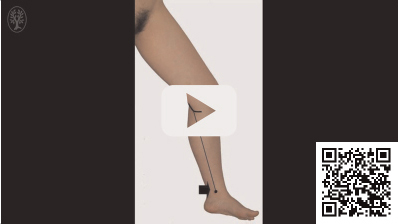
Video 15.1 Island Flap: Medial Sural Artery Perforator Flap. https://www.thieme.de/de/q.htm?p=opn/cs/20/7/12265268-f12f31bb
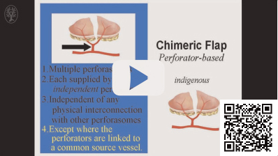
Video 15.2 Island Flap: ALT Chimeric Perforator Flap. https://www.thieme.de/de/q.htm?p=opn/cs/20/7/12265269-459e13e0
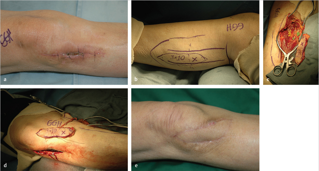
Fig. 15.4 (a) Dehisced wound after right total knee replacement with quadriceps tendon exposure. (b) Following debridement, a medial sural artery perforator (MSAP) island flap was designed over the outlined medial gastrocnemius (MG) muscle, intentionally eccentric distal to a perforator identified with audible Doppler marked “x.” (c) Perforator dissected through the MG muscle back to a main medial sural artery branch (green microgrid) to extend the flap vascular leash. (d) Reach of this island MSAP flap seen before passing through a shorter pathway via a subcutaneous tunnel to the defect, (e) allowing tension free healing.
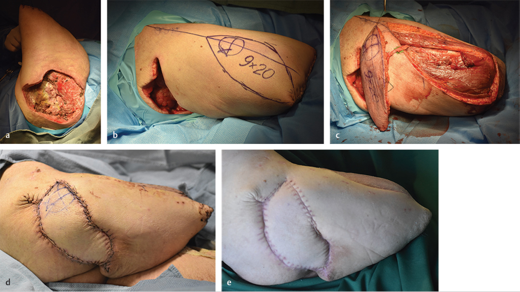
Fig. 15.5 (a) Necrotic right hip wound, with right above-knee amputation after frostbite. (b) Following serial wound debridement, planned anterolateral thigh (ALT) island flap designed as eccentrically as possible toward stump distal to perforator “x” to maximize flap reach. (c) Proximal-pedicled (on microgrid) ALT flap transposed toward right hip. (d) Flap inset after it is drawn through the subcutaneous tunnel between the thigh donor site and the right hip. (e) Right hip healing after 5 weeks, as is skin graft necessary to close the donor site.
15.2.7 Postoperative Care Protocols
The potential for compression not just of the vascular pedicle to these island flaps that are usually passed through a subcutaneous or submuscular tunnel, but also the diminutive perforators themselves requires that the flaps be observed as carefully as per the usual protocol for a free flap. If the flap crosses a joint, immobilization is essential to prevent movement resulting in stretching of the pedicle, its obstruction or disruption, or even flap dehiscence.
15.2.8 Conclusion
A “true” island perforator flap not only will have all its skin boundaries disconnected from those of its donor site but also will have an elongated pedicle extending beyond just the flap perforator itself, or else would more properly be called an “islanded” flap. This will therefore require inclusion of a major branch or the source vessel of the perforator itself as part of the flap pedicle. Previously, this has been accomplished by sacrificing either the anterior tibial,5 posterior tibial,6 or peroneal7 artery, but this is no longer an acceptable risk under any conditions for fear of eventual devascularization or ischemia of the foot. Other more straightforward local perforator flap alternatives today have become superior options.16 However, the territories of the ALT or MSAP flaps still represent pragmatic choices for island flaps that as such have far greater reach for coverage of proximal or distal defects of the lower limb than do other local perforator flap subtypes.
References
[4] Behan FC. The keystone design perforator island flap in reconstructive surgery. ANZ J Surg. 2003; 73(3):112–120
15.3 Chapter 15B: Perforator Advancement Flaps Including the Keystone Flap
Geoffrey G. Hallock
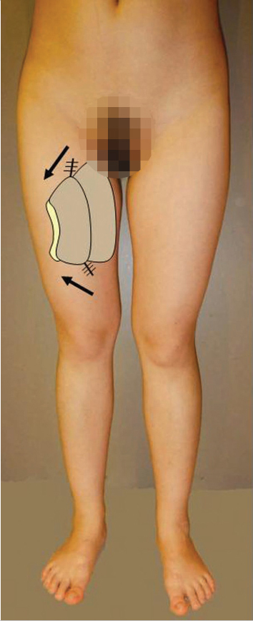
Fig. 15.6 The Keystone Advancement Flap.
15.3.1 Introduction to the Perforator Advancement Flaps Including the Keystone Flap
An advancement flap by definition is always moved in a forward direction into a defect without any rotation or lateral movement.1 A classic prototype was a unipedicled quadrilateral-shaped random flap sustained by a skin bridge on one side that is now primarily only of historical interest as this geometric arrangement has limited mobility and therefore inadequate reach in the lower extremity.1 The advent of the perforator flap concept provided an improved means for advancement flap vascularization, but practically has been limited in the lower extremity to V-Y advancement and keystone island flap designs.
According to Niranjan et al,2 Blasius (1848) should be credited with the idea of the V-Y advancement flap, which for a long time was based on a subcutaneous pedicle3 with reach dependent on the sliding capability of these triangular shaped skin flaps.4,5 Since in actuality the fascial plexus alone within the subcutaneous tissues can be quite tenuous, these flaps more likely have always received their circulation from unrecognized perforators of the deep fascia and fascia feeders surrounding it.2 The same comments also apply to the recent variation introduced by Behan6 as the “keystone design perforator island flap (▶ Fig. 15.6).” This basically is a trapezoidal islanded flap7 that consists of two conjoined7,8 or opposed9 V-Y advancement flaps. Although usually stated to be islanded, the keystone flap still maintains some connection to the deep fascia directly underneath it. In contradistinction, additional reach no longer relying on skin laxity of a V-Y advancement flap is possible if the flap can be isolated on a distinct perforator,2,7,10,11 whereas the multiple perforators to a keystone flap are never specifically sought out.12
15.3.2 Attributes and Detriments
Attributes
• Local flaps adjacent to defect.
• Similar texture and contour.
• Design simplicity.
• Rapid elevation.
• Nonmicrovascular tissue transfers.
• Minimal donor site morbidity.
Detriments
• Often require adequate soft-tissue laxity for advancement.
• V-Y advancement flap limited to smaller defects.
• Although gigantic keystone flaps are possible, significant scar residue is a sequela.
• If deep fascia cannot be readily separated, these may not easily span space across bone, joint, or prosthesis.
• Lower extremity defects are more likely to require circumferential deep fascia release for keystone flaps.
15.3.3 Anatomical Considerations
Neither a V-Y advancement flap nor a keystone flap absolutely requires identification of a specific perforator. Both can be supplied by perforators of the deep fascia, which could be no more than the ubiquitous capillary perforator,13 or fascia feeders.2 As such, these flaps should be oriented to overlie known perforators or areas of high perforator density, that is, “hot spots,”12 following the longitudinal course of the three source vessels to the foot. If a perforator can be precisely determined preoperatively using available adjunctive techniques, a V-Y advancement flap could instead become an islanded flap connected only to that specific perforator, which would thereby have farther reach, albeit still limited by the length of the perforator itself.
As with any local perforator flap, the condition of local tissues and possible compromise of perforators must always be assessed carefully. In addition, unique to the lower extremity, advancement flaps may be more difficult,14 as tissue laxity or slipperiness may be nonexistent over the deep fascia although still probably adequate over the muscles of the thigh and calf, if anywhere. As a consequence, transverse oriented V-Y advancement flaps are more mobile,5,11 as would be longitudinally designed keystone flaps.9 For keystone flaps, this is an advantage, as this best recruits circulation from multiple captured perforators perhaps from adjacent territories according to the perforasome theory.15
15.3.4 Anatomical Variations and Potential Pitfalls
Extended Reach
Salvage of Intrinsic Flap Tissues
Rather than discarding the tissues of the margins of the leading edge of an advancement flap, these can be sewn together, thereby increasing the width of the flap and consequently lessening the amount of advancement needed. For the V-Y advancement flap, this maneuver has been called the PAC-Man technique2,16,17 (▶ Fig. 15.7) and for the keystone flap, the omega technique14 (▶ Fig. 15.8).
Deep Fascia Release
Often an incision of the deep fascia as needed about either the V-Y advancement or keystone flap will allow greater movement, as restraints from connections to the underlying tissues are partially released. Usually this begins at the greater curvature border of the keystone flap9,12 and progresses outward as required to obtain the desired flap mobility. Subfascial extension of this dissection to get even more release should best be away from areas of known high perforator density so they will not be injured.
Suprafascial Flap Release
Instead of incising the deep fascia and violating perforators and perhaps fascial feeders, a suprafascial dissection raising the subcutaneous tissues of either the V-Y advancement or keystone flap from the deep fascia as needed for further advancement can be done. This requires careful preservation of any small vessels encountered, which makes this a more difficult alternative as microdissection will be required.18 The V-Y advancement flap should be elevated at its apex and base first, whereas for the keystone flap, margins should be explored where perforator density is known to be the least.
Rotation Advancement Flap
If an islanded V-Y advancement flap is intended, but during the subfascial dissection through the exploratory incision via one side of the flap no adequate perforator is found, the flap should be redesigned retaining the contralateral side intact. The base can then be advanced forward with some rotation about its intersection and the retained side to then close the defect19 (▶ Fig. 15.9). This may require a small back-cut of the retained side starting at the flap apex to allow adequate rotation.
Keystone “Plus” Flap Design
A “V”-shaped extension half of the defect width is designed in the center of the greater curvature of the arc that connected the tangential wings of the standard keystone flap (▶ Fig. 15.10).20 After flap advancement, this donor site is also closed in a V-Y fashion, which should relieve tension on closure of the greater curvature side of the overall donor defect.
Keystone Flap Skin Bridge Design
The cutaneous central member of the greater arc of the keystone flap is not incised, although the deep fascia is released in its entirety underneath this skin bridge that has been left intact (▶ Fig. 15.11). This should not restrict closure of the donor site. Subdermal lymphatics from the flap are preserved, and Moncrieff et al21 believe the flap will also have greater vascularity via this connection. However, this contradicts the opinion of Behan14,22 who believes that the creation of a completely islanded flap causes a sympathectomy effect that results in augmented, not diminished, flap perfusion.
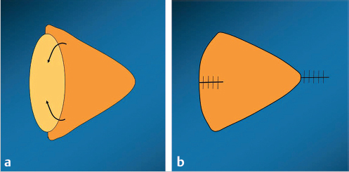
Fig. 15.7 (a) PAC-Man technique: sides of base of V-Y advancement flap (orange) are sewn together in the direction of the arrows, (b) which lengthens flap, reduces the needed advancement, and otherwise is inset in a V-Y fashion.
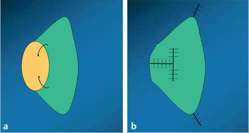
Fig. 15.8 (a) Omega technique: sides of lesser curvature of keystone flap (green) are brought together in the direction of the arrows and (b) then sewn to each other and to the middle of the lesser curvature to form a resemblance of the Greek letter “Ω” that increases the overall width of the flap.
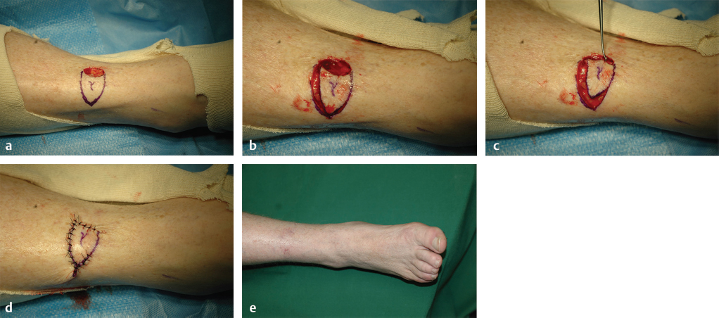
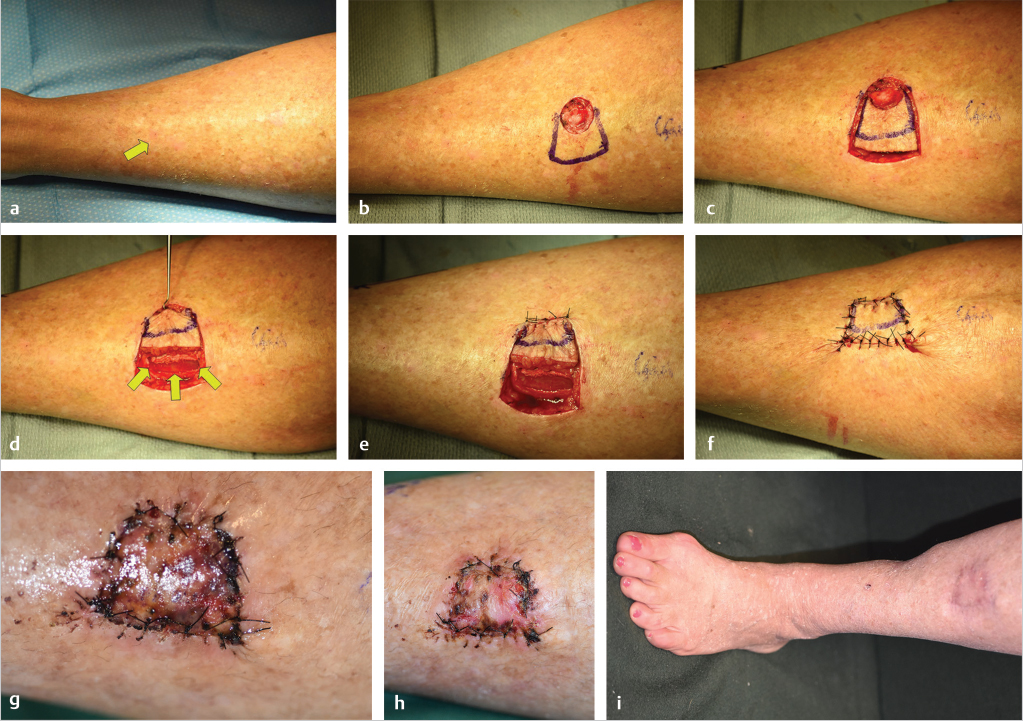
Fig. 15.9 (a) Anterior right distal leg skin cancer excision, with proposed transverse oriented V-Y advancement flap for closure about perforator marked “y.” (b) Subfascial exploratory incision through one side of the triangle did not reveal a perforator, (c) so the other side of the flap was left intact except for short back-cut that facilitated flap rotation and advancement to reach the defect. (d) Completed flap insetting with direct donor site closure. (e) Ultimate superior appearance of the leg without a skin graft.
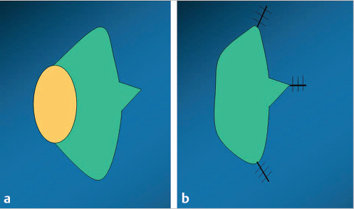
Fig. 15.10 (a) Keystone plus flap: “V” extension at the center of the greater curvature of the keystone flap (green) is intended to decrease tension on the latter’s closure after advancement, (b) as that extension donor site will be closed to gain the same advantage as a V-Y advancement.
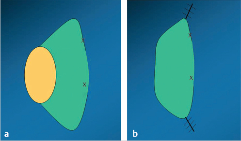
Fig. 15.11 (a) Keystone bridge flap: skin of the central portion of the greater curvature (area between “Xs”) of the keystone flap (green) is not incised and (b) always remains attached to the advanced flap when inset as well as at the original donor site.
15.3.5 Flap Design
V-Y Advancement
The laxity of tissues adjacent to the defect will determine the orientation or even possibility of using this option as a subcutaneous pedicled flap as the keystone flap is typically used. The base of the isosceles triangular shaped flap should slightly exceed the width of the defect (▶ Fig. 15.12). The altitude of the flap will extend perpendicular to the base up to 1.5 to 2.0 times the height of the defect, with a longer flap probably allowing easier closure of the donor site at the apex of the flap that will be in the vicinity of the most pliable donor site tissues.
If subcutaneous tissue elasticity is known to be inadequate for the needed advancement, preoperative search for a nearby perforator should be undertaken. If the perforator is close to the defect, a propeller flap may be a preferred option.7 If the perforator is further from the defect, it may be more appropriate to design a V-Y advancement flap using the same dimensions as for the subcutaneous pedicled version, although now instead centered over the requisite perforator (▶ Fig. 15.13).
Keystone Flap
Behan classified keystone flaps into four types that varied by the degree of undermining of the deep fascia, number of flaps used, or treatment of the donor site, which rarely required a skin graft.6 His standard format is a representative model of all his subtypes that will be the focus here (▶ Fig. 15.14). Although in the past many have extended the original defect in an elliptical fashion to coincide with the lesser curvature of the flap,14 in the lower extremity this can be a waste of precious normal tissues that may instead require alteration of the flap design (▶ Fig. 15.15).8 Local tissues about the defect should be assessed for the presence of perforators and compared to areas with maximum skin laxity. This can be facilitated by the use of an audible Doppler; however, knowledge of known high-density perforator locations, that is, “hot spots,”12 or the fact that perforators commonly emanate following the longitudinal course of the source vessels to the foot can be used safely with impunity. Since perforasomes in the lower extremity interconnect in a longitudinal direction,15 the keystone flap should have a similar axis especially if the most pliable tissues can be simultaneously captured.
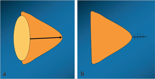
Fig. 15.12 (a) Basic design of V-Y advancement flap (orange) as a triangle with the base slightly greater than the width of the defect (yellow), and altitude (perpendicular line to the apex) 1.5 to 2.0 times the height of the defect itself. (b) After advancement to close the defect, the donor area behind the apex is closed primarily to result in the ultimate “Y” appearance of the overall closure.
Wings starting tangential to the defect on opposite sides should proceed at about a 90-degree angle away for at least as far as the height of the defect. This distance determines the width of the keystone flap. The two tangent lines are joined by an arc to complete the trapezoidal or keystone shape of the flap. If the lesser curvature edge of the flap has already been undermined, its tissues are of poor quality such as irradiated, inelastic, or traumatized, the wings and thus the width of the flap can be increased even up to 5:1 the size of the defect so as to capture adequately perfused and elastic tissues having the desired qualities that can then be advanced as needed.12
15.3.6 Flap Harvest
The position of the patient on the operating room table will depend on the location of the defect and the proposed design of the local perforator flap chosen for its correction, so as to most simply allow access to both.
V-Y Advancement
If subcutaneous tissue laxity is readily mobile, the sides of the designed triangle are incised down to at least the equivalent of Scarpa’s fascia, or to the deep fascia itself if necessary. Often, the subcutaneous tissues at the base of the flap can be carefully raised from the deep fascia as is also done at the apex, as far as required, to then allow additional reach so that the base can be sewn to the farthest edge of the defect. This dissection requires preservation of any small vessels encountered as would be done routinely in the raising of a perforator flap. The triangle’s sides are sewn where advanced to the corresponding sides of the defect and donor site. The donor site inferior to the flap apex can then be closed primarily to complete the repair and “Y” appearance of the closure.
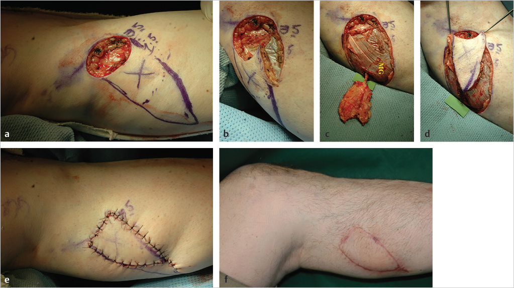
Fig. 15.13 (a) Following excision of a basal cell carcinoma from the right calf, a propeller flap was initially planned on a medial sural artery perforator (MSAP) “x” heard with the audible Doppler, (b) yet the obligatory exploratory incision revealed that the perforator instead was near the apex of the flap, so a V-Y advancement flap design became a more reasonable alternative. (c) Enough reach required some intramuscular dissection of that perforator (microgrid) into the medial gastrocnemius muscle (MG), (d) sufficient so that the required tension-free advancement of this MSAP V-Y advancement flap was possible, (e) allowing insetting for closure of the defect and donor site in a “Y” fashion, (f) with result 3 months later where a skin graft had been avoided.
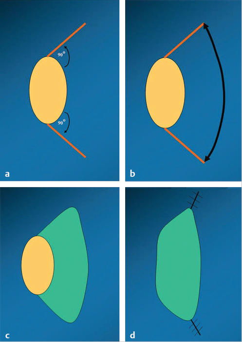
Fig. 15.14 (a) Standard keystone flap: tangents on opposite sides of the defect (yellow) are drawn at about a 90-degree angle, with length at least equal to the defect width, (b) an arc connects the ends of the tangents (c) to form a trapezoid- or keystone-shaped flap (green) (d) that will then be advanced to close the defect, with the small open areas remaining on either side of the greater curvature closed in a V-Y fashion.
If the initial plan is to seek a preoperatively identified subfascial perforator that will better allow advancement of an islanded V-Y advancement flap, only one side of the triangle is incised through the deep fascia as an exploratory incision. The perforator is isolated and lengthened as necessary by coagulation of side branches back to its source vessel or branch. If found adequate, the other flap side is then completely incised, and the flap advanced without tension for insetting just as was done for the subcutaneous pedicled version. However, if no adequate subfascial perforator is confirmed, the backup option of a rotation advancement flap must be considered (▶ Fig. 15.9).19

Video 15.3 Keystone Advancement flap. https://www.thieme.de/de/q.htm?p=opn/cs/20/7/12265270-583aca14
Keystone Flap
The wings and greater arc of the standard designed keystone flap are incised (see ▶ Video 15.3). This is carried down to Scarpa’s fascia or its equivalent first; if flap advancement is easily possible, some portion of the subcutaneous fascial plexus from tissues adjacent to the flap itself can be preserved for vascular augmentation. However, if necessary, a stepwise release even of the deep fascia starting at the midpoint of the outer curvature incision, then even circumferentially about the flap may have to be done until the flap can be advanced for closure without tension.9,12 In addition, this could require even some subfascial dissection for further release from underlying tissues, but always away from the known location of flap perforators. Once satisfactory, the center of the flap’s inner curvature border is advanced to the middle of the farthest edge of the defect, where subcutaneous sutures are placed to reinforce the attachment. The skin there is next closed as are the sides of the flap, with trimming of any excess tiny remnants at the leading edge of the flap. Only the skin of the outer curvature line is then reclosed in a relatively loose fashion. This will leave a short open area on either side of the arc of the flap, which is closed in the typical V-Y flap fashion to complete the reconstruction.
15.3.7 Postoperative Care Protocols
Both the V-Y and keystone advancement flap versions pedicled by their deep fascial attachments are extremely robust flaps requiring little additional scrutiny as long as pressure is kept off them and adjacent joints relatively immobilized to minimize compression and stretch. On the contrary, an islanded V-Y advancement flap must be carefully protected so that the perforator itself will not be compromised, by appropriate dressings and positioning of the patient, for at least a week or more in most patients until wound healing stability is satisfactory. Sutures, especially in the keystone flap, should never be removed in a hurry, perhaps waiting several weeks or more sometimes so that dehiscence does not occur.
15.3.8 Conclusion
Local perforator advancement flaps basically allow only forward movement of tissues adjacent to a given defect. In the lower extremity, the most pragmatic variations are limited to the V-Y advancement or keystone flap. These on a geometric basis have great similarity in design, and also both can survive on multiple perforators through the deep fascia without the need for specific perforator identification. Yet in this form, both rely on tissue laxity to allow the required advancement, with the keystone flap concept necessitating at times even gigantic flaps if need be to obtain this reach. The V-Y advancement flap overall is limited to small or moderate-sized defects, where any additional reach if desired requires instead that a specific perforator be found so that an islanded version tethered only by the length of that as the vascular pedicle will prove to be sufficient.
Related posts:
 General Wound Preparation and Timing
 The Pertinence of the Reconstructive Ladder and the Reconstructive Elevator
 Supermicrosurgery Approach to the Lower Limb
 Nonflap Wound Closure Alternatives: Skin Graft, Skin Substitute, Skin Stretch, and Negative-Pressure Wound Therapy
 Vascular Anatomy of the Lower Extremity: A Practical Guide to Vascular Territories, Perforators, and Selection of Recipient Vessels
 Using the Flap and Angiosome Concepts to Optimize Functional Lower Leg and Foot Amputations
Stay updated, free articles. Join our Telegram channel

Full access? Get Clinical Tree


