17 Supermicrosurgery Approach to the Lower Limb
Summary
Supermicrosurgery is manipulating vessels with diameters less than 0.8 mm. In flap surgery, this approach will allow an increase in the selection of recipient pedicles by using a perforator-to-perforator anastomosis approach. Decreased time of surgery, elevation of the flap by taking just a short segment of the perforator pedicle, minimization of any risk for major vessel injury, and utilizing collateral circulation in ischemic diabetic foot are some of the apparent advantages.
Keywords: supermicrosurgery, perforator-to-perforator microsurgery, diabetic foot reconstruction, freestyle reconstruction
17.1 Introduction
Reconstruction of the lower extremity using free flaps remains a difficult procedure. Although limb salvage rates have greatly improved over the past 20 to 30 years, major hurdles regarding single artery limb, atherosclerosis, involvement of hardware, frequent spasm, chronic infections, edema, complexity of lower limb trauma, and diabetes still make it very challenging even for experienced surgeons.1 In this age of reconstruction, we are faced with the challenge of achieving not only successful soft-tissue coverage but also functional recovery, infection control, and cosmetic improvement. Evolutions in microsurgery have been noted from the days of experimental surgery, finger replantation, and now to perforator flaps and supermicrosurgery, and it has allowed achieving complex goals. These evolutions aim for better results and to minimize complications despite the innate risks of lower extremity soft-tissue reconstruction. The use of perforator-to-perforator supermicrosurgery has reduced complications in high-risk lower extremity reconstructions.
Lower extremity can be extremely difficult to reconstruct with poor outcome. Frequently the lower extremity reconstruction involves major vessel injury leaving only one major vessel to the leg, requires long pedicle length to reach the recipient vessel, may have severe arthrosclerosis in diabetic or elderly patients limiting the choices for recipient vessels, has lower perfusion, being furthest from the heart, and is very difficult to isolate the recipient vessels in the mid and upper leg as it is lying very deep to the skin. How can we overcome these difficult situations without compromising the outcome of reconstruction while maintaining a robust distal flow? This is the key challenge.
The supermicrosurgery technique is defined as microsurgical anastomosis of vessels, with a diameter less than 0.8 mm.2,3,4,5 This technique, although reported frequently on lymphaticovenous shunting to treat lymphedema, fingertip replantation, finger/toe reconstruction, and sporadically in soft-tissue reconstruction with specific indications, is a relatively new concept for lower extremity reconstruction.3,6,7,8,9,10,11,12,13 As lymphedema supermicrosurgery will be covered in another chapter, this chapter will focus on soft-tissue reconstruction. For the lower extremity soft-tissue reconstruction, one of the applications can be seen in the perforator-to-perforator anastomosis approach or using small-diameter pedicles of perforator flaps to anastomose on a lager vessel end to side.4,5,14,15 This concept deserves to be recognized as a new paradigm in microsurgery as it has been mentioned that vessel size less than 1 mm may significantly increase the risk of flap failure.16 However, with advances in technology and skills, the overall success rate for using supermicrosurgery technique for lower extremity reconstruction compares similar to other reports using vessels larger than 1 mm.2,16,17,18
The evolution into this new concept and paradigm shift came naturally as the understanding of single perforasome (perforator angiosome) unfolded as well as the establishment of free styling of perforator flap elevation.19,20,21,22,23,24 The successful survival of the skin flap based on a single perforator led the hypothesis of using a single perforator artery and veins as a recipient pedicle.4,5,14 Thus, using the perforator as a recipient vessel may overcome the difficult challenges that the surgeons face during lower extremity reconstruction. Introduction of supermicrosurgery, especially perforator to perforator, also led to true free style free flaps as any perforator-based skin flaps even the one with short pedicles now can be used to reconstruct the defect.4,10,14,25,26,27
Another possibility that perforator-to-perforator supermicrosurgery showed was in treating ischemic diabetic foot patients. Patient with ischemic diabetic limb develop multiple collaterals as atherosclerosis develops. It is these collateral vessels that can supply the skin perforators to maintain the vascular flow despite of major artery obstruction. Using these collateral arteries and perforators supplied from these collaterals is essential for the application of supermicrosurgery in ischemic diabetic limb.15,28,29 The details of this approach is described in the chapter for diabetic foot reconstruction.
In this chapter, we will focus on how the perforator-to-perforator supermicrosurgery can help surgeons to overcome some of the challenges seen in lower extremity reconstruction.
17.2 Attributes and Detriments
17.2.1 Attributes
• Perforators or small end vessels can be used as recipient vessels.
• Perforator-to-perforator supermicrosurgery alleviates the use of major vessels and maintains the distal flow to the leg without disruption.
• Pulsatile perforator or an end vessel is a good indication for use.
• This approach can be minimally invasive, thus minimizing the need for extensive dissection to isolate the recipient.
17.2.2 Detriments
• Special instruments may be required for supermicrosurgery.
• There is a steep learning curve.
• Perforator as a recipient has a limit in providing perfusion to the tissue.
• Volume depletion must be avoided during post-op care.
17.3 Anatomical Consideration
There are more than 420 perforators that are over 0.5 mm in diameter throughout our body.30 While these perforators can be seen as potential sites for perforator-based flaps, they can be considered as recipient vessels as well. In the lower extremity, there are on average 184 perforators from gluteal, hip, thigh, knee, leg, and foot.30 The abundance of these potential recipient sites truly allows us to approach in free style in not only elevating the flaps, but also selecting the recipient source.4,5 The perforator is an end vessel that is the sum of all supplies not only from major vessels but also from all the branches and collaterals heading toward the end vessel perforator.15,28 Thus, using these perforator or end vessels becomes possible despite the major artery being insufficient from atherosclerosis, trauma, or other relevant causes.4 Even in legs with single artery, the risk of steal phenomenon or distal flow reduction can be avoided as using perforators will not disturb the flow of this major vessel.
Preoperative CT scans will allow us to view the vascular details of the lower extremity. In the cases where foreign body effect deters from obtaining appropriate images, conventional angiograms can be obtained as well. These images can also show the possible recipient sites as well as give information on potential donor flap sites. Preoperative duplex can be used to enforce the findings from Doppler as this can give you objective measurement such as velocity of flow and the diameter of the vessel as well as more detailed anatomical findings.
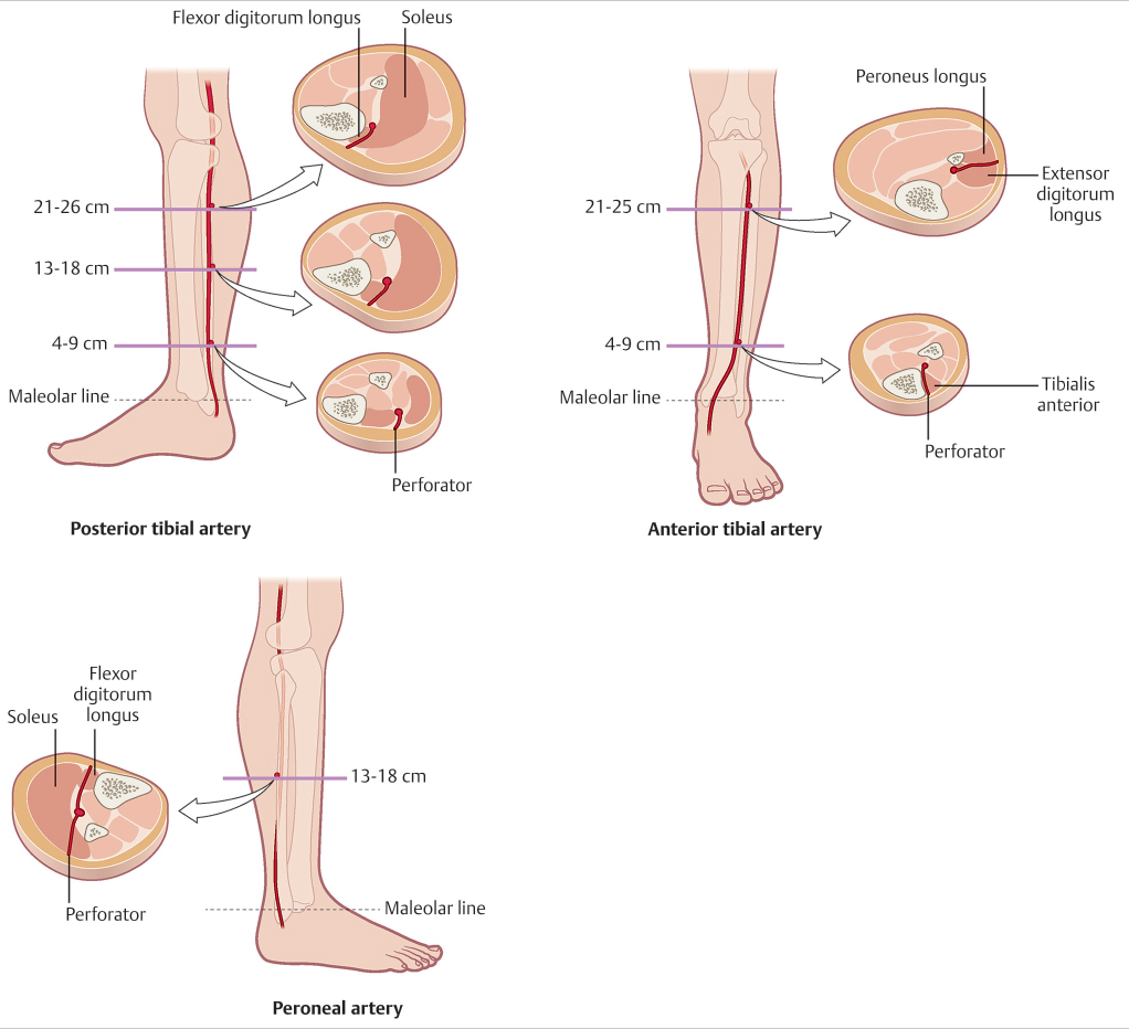
In the lower extremity, there are relatively constant and large perforators coming out from the three major vessels. The findings of the study by Schaverien and Saint-Cyr are shown in ▶ Fig. 17.1, which depicts the relatively constant regions where the perforator emerges from the three major arteries.31 The perforators from the posterior tibial artery are found between the flexor digitorum longus and soleus in all three intervals, with the largest perforators found in the middle cluster. The constant perforators from the anterior tibial artery are located within two clusters. The proximal perforators, 21 to 25 cm from the intermalleolar line, are in the anterior peroneal septum between the extensor digitorum longus and the peroneus longus, and the distal perforators, 4 to 9 cm from the intermalleolar line, are found between the tibia and the tendon of the tibialis anterior muscle. The constant perforator for the peroneal artery is found 13 and 18 cm from the intermalleolar line emerging from various muscles.31 Using these relatively constantly located perforators will allow the surgeon to approach with some degree of reliability when considering the perforator as a recipient.
Related posts:
 General Wound Preparation and Timing
 The Pertinence of the Reconstructive Ladder and the Reconstructive Elevator
 Modern Concepts of Prosthetic Rehabilitation in Amputation of the Lower Extremity
 Lower Limb Vascularized Composite Allotransplantation
 Using the Flap and Angiosome Concepts to Optimize Functional Lower Leg and Foot Amputations
 Procurement of Thin Flaps as Indicated in the Lower Extremity
Stay updated, free articles. Join our Telegram channel

Full access? Get Clinical Tree


