10 General Wound Preparation and Timing
Summary
With few exceptions, the lower extremity defect is rarely suitable for reconstruction at the time of initial presentation. Preoperative patient optimization such as by smoking cessation, assurance of adequate nutrition, diabetes management with glucose control, and correction of any vascular insufficiencies will allow superior wound healing and lessen the risk of complications. The timing of wound closure is then controversial, but preferably before the onset of the subacute phase and definitely not until initial irrigation to remove contaminants and lessen any risk of infection, followed by serial debridement that removes all nonviable elements and residual biofilm, has been completed.
Keywords: nutrition, debridement, biofilm, timing of wound closure
10.1 Introduction
Lower extremity reconstruction can be necessary for multiple reasons, ranging from traumatic injury to oncologic extirpation to infection and osteomyelitis. Except for immediate reconstruction after tumor resection, the wound is rarely ready for the final repair at the index presentation. Preparation of the wound and timing of the reconstruction are two variables of paramount importance to consider for optimizing outcomes.
10.2 Patient Optimization
Whenever possible, proper patient optimization can have a substantial positive impact on the outcome in the elective setting. For example, smoking is well known to be detrimental to wound healing and tissue vascularity.1,2 Preoperative and postoperative smoking cessation has been shown to reduce surgical complication rates.3 Patients should abstain from nicotine use for at least 4 weeks before and 4 weeks after any elective surgery in order to reduce complication rates to the lowest possible level, although it is never superior to that of the nonsmoker.2
Nutrition is also a critical consideration, as protein deficiency, particularly deficiencies of arginine and methionine, is known to have a substantial deleterious impact on wound healing. Preoperative nutritional support has been shown to reduce the rates of infectious and noninfectious surgical complications.4,5 Therefore, serum nutritional markers should be checked and optimized preoperatively, with a goal for a serum albumin greater than 3.25 g/dL, and serum prealbumin greater than 15 mg/dL.2
In diabetic patients with lower extremity ulcers, perioperative glucose control is of paramount importance. The risk of surgical-site infections increases proportionately to serum glucose above 110 mg/dL,6 and even a single perioperative instance of serum glucose greater than 200 mg/dL has been found to significantly increase the risk of wound dehiscence.7 Hemoglobin A1c should be ≤ 7.5% before any elective surgery is undertaken to optimize outcomes.2
Patients with compromised vascular inflow into the lower extremity, whether due to medical comorbidities or trauma, must have a vascular profile of that limb meticulously evaluated and optimized before reconstruction is attempted. The potential for healing is enhanced when the periwound transcutaneous partial pressure of oxygen is 40 mm Hg or greater.8
Prior to any reconstruction in the trauma patient, stability of homeostatic mechanisms always takes precedence. A full standard trauma survey of all systems must be completed, and then repeated routinely. If lower limb vascular injuries are identified, a preoperative angiogram can provide invaluable diagnostic data, and ascertain the need for vascular surgery intervention. Depending on the circumstances and risk of a compartment syndrome, fasciotomies may be required. Fractures must next be reduced and stabilized. Finally, thorough irrigation and debridement of the wound bed is a fundamental and critical cornerstone for success. The ultimate choice of reconstructive modality must take into account the exposure of vital structures, such as tendon, bone, nerve, and blood vessel.
10.3 Timing of Lower Extremity Coverage after Trauma
Timing of posttraumatic lower extremity reconstruction has been debated in the literature for decades. A landmark study by Godina in 1986 dramatically influenced the timing of posttraumatic lower extremity reconstruction in open tibia–fibula fractures.9 In this study, 532 patients were categorized into three groups based on timing of free flap reconstruction after trauma: less than 72 hours (acute), 72 hours to 3 months (subacute), and greater than 3 months (chronic). Godina found that coverage in the acute phase resulted in the lowest rates of postoperative infection, flap failure, bone healing time, and hospital length of stay. Coverage in the subacute period resulted in the worst outcomes. As a result, early flap coverage of open tibia–fibula fractures within 72 hours of injury has generally been proposed as the ideal time for reconstruction.
Similarly, Byrd et al emphasized the importance of thorough debridement and acute (defined as within 5 days) microsurgical coverage of open tibia–fibula fractures.10 They classified open tibial fractures based on the amount of energy involved in the trauma, the degree of bone comminution, and the extent of soft-tissue injury. Higher-grade injuries were characterized by greater bone fragmentation and bacterial colonization, and thus had worse outcomes.
Other studies have sought to challenge the acute coverage paradigm. Francel et al argued that good surgical and functional outcomes are achievable when reconstruction is performed up to 15 days after Gustilo grade IIIB tibia–fibula fractures.11 However, these good outcomes are predicated on meticulous wound debridement with excision of any necrotic tissue or debris, and serial washouts. Emphasis was placed on debridement of devitalized bone, which may otherwise form a sequestrum and subsequent nidus for infection. Recent military experience has also demonstrated that delaying reconstruction to the subacute time period, with intervening serial debridements, has an acceptable complication rate for reconstruction.12,13,14
Negative-pressure wound therapy (NPWT) is one of the advancements that have facilitated delayed lower extremity reconstruction. NPWT has been shown to promote granulation tissue formation, enhance wound contraction,15,16,17 remove inflammatory cytokines and metalloproteinases,18 and decrease bacterial burden.19 By delaying reconstruction with the use of NPWT, tissue edema can decrease, and areas of necrosis are allowed to demarcate.13,20 NPWT therefore serves as a valuable dressing between serial debridements, but always in the preparation for definitive reconstruction. A novel advance in NPWT is the addition of instillation therapy, whereby the negative suction is intermittently interrupted for a few minutes, and saline or a topical antiseptic is instilled into the wound, allowed to dwell, and then suctioned out. NPWT with instillation significantly reduces wound bacterial contamination.21,22
At the time of serial debridement, the surgeon should obtain quantitative bacterial tissue culture. A positive quantitative culture may warrant further debridement before reconstruction, as reconstruction in the setting of a positive culture has been shown to portend significantly higher complication rates.23
10.4 Wound Bed Preparation
Wound bed preparation is the most critical step to ensure successful lower extremity reconstruction. Multiple methods of cleansing wounds exist, but the fundamental principles of removing foreign bodies and debriding devitalized tissue are the ultimate focus of all methods of wound bed preparation.
Irrigation is the initial step in cleansing a contaminated or infected wound. It allows for the simple removal of foreign bodies and contaminated tissue. Normal saline is the most common solution used, and there is little evidence to suggest any sustained benefit to additives such as antibiotics or detergents.24 The irrigation may be delivered to the wound under low pressure (1–15 psi), such as cystoscopy tubing, or under high pressure, such as pulsed lavage (35–70 psi).25 Some literature has suggested that high-pressure irrigation may be more effective at reducing bacterial counts and wound infection rates.26,27 However, there is also concern that high-pressure irrigation causes soft-tissue damage28 and bacterial propagation into deeper tissues, such as bone.29
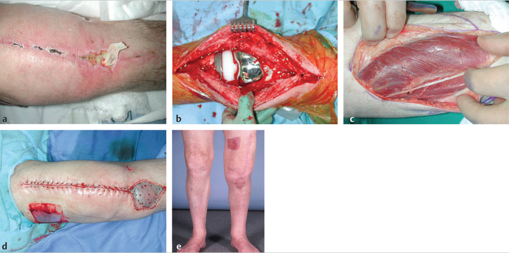
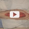
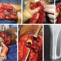
The second step in wound preparation is complete debridement of any necrotic tissue. The goal of debridement is to achieve residual tissues that are clean, viable, and free from contamination (▶ Fig. 10.1). A goal of debridement is also to address and remove bacterial biofilm. Bacteria encased in biofilm are especially resilient even in the face of antibiotic therapy.30 Surgical debridement temporarily converts bacteria in the biofilm to a planktonic state, rendering them more susceptible to antimicrobial treatment.
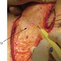
Surgeons who are comfortable with reconstruction may be more aggressive at making a wound larger through debridement, knowing that they will be able to cover the resulting defect. In the traumatic wound, sharp excisional debridement is the most commonly used modality. This can be performed with conventional surgical instruments, such as scalpels, scissors, and curettes. Debridement can also be achieved with hydrosurgery (Versajet, Smith & Nephew, Cambridge, UK), which uses a high-speed stream of water to generate a localized vacuum via the Venturi effect (▶ Fig. 10.2), with the subsequent removal of nonadherent tissue.31,32 Hydrosurgery has been found to be more efficient and result in less blood loss than conventional sharp debridement.33 It also has been shown to more effectively address biofilm than mechanical debridement alone34 or high-pressure pulsed lavage.35
Related posts:
 Indications for Vascular Intervention in Lower Extremity Reconstruction
 Supermicrosurgery Approach to the Lower Limb
 Modern Concepts of Prosthetic Rehabilitation in Amputation of the Lower Extremity
 Lower Limb Vascularized Composite Allotransplantation
 Using the Flap and Angiosome Concepts to Optimize Functional Lower Leg and Foot Amputations
 Procurement of Thin Flaps as Indicated in the Lower Extremity
Stay updated, free articles. Join our Telegram channel

Full access? Get Clinical Tree


