3 Indications for Vascular Intervention in Lower Extremity Reconstruction
Summary
Proper initial assessment of the peripheral vascular status of the impaired lower limb should never be overlooked, as a balance between the arterial and venous systems is essential for equilibrium of the homeostatic mechanisms responsible for tissue regeneration and wound healing. Vascular laboratory evaluations that include basic ankle-brachial indices and pulse volume recordings, as well as standard imaging technologies, may be required for staging and characterizing the extent of any disease processes. These will aid in determining if a vascular surgery consultation would be essential, often mandatory before any reconstructive endeavor.
Keywords: peripheral vascular disease, ankle-brachial index, pulse volume recordings
3.1 Introduction
Uncomplicated lower extremity wound healing relies on adequate tissue perfusion. Satisfactory arterial inflow allows access for immune cells and provides metabolic factors for the inflammatory process and the subsequent anabolic stages of healing. Likewise, venous outflow will provide for the elimination of undesirable metabolites and byproducts. This important balance underlies the homeostatic mechanisms of wound healing and tissue remodeling; likewise, disturbances in this perfusion–drainage equilibrium will contribute to pathologic changes in the healing process and may cause de novo wounds to develop from ischemia, venous stasis, or a combination of the two.
The lower extremity is at increased risk for peripheral vascular disease (PVD) and often presents a significant challenge for the reconstructive surgeon. Patients with known peripheral arterial disease (PAD) or the stigmata thereof (i.e., claudication, ulceration) and those with risk factors for atherosclerotic disease (smoking, obesity, diabetes) must be carefully evaluated and screened. Failure to first appreciate vascular disease is the cardinal sin of lower extremity reconstruction as it will lead to catastrophic outcomes.
The progress realized in reconstructive surgery over the past three decades has been paralleled by our vascular surgery colleagues. In the 1970s, aortofemoral bypass and/or profundaplasty were the only vascular surgical options to improve the blood supply to a chronically ischemic limb. Today, bypass grafting and endovascular procedures below the popliteal artery, and all the way into the foot are commonplace.
3.2 Principles of Limb Salvage
Successful limb salvage or reconstruction depends on proper patient selection, judicious preoperative preparation, and meticulous postoperative care. Anyone embarking upon a career in the management of lower extremity wounds must understand the relationship between the reconstructive options conferred by the phasic nature of blood flow into the limb and the amount of tissue required for the reconstructive effort. It is critically important that the reconstructive surgeon is well versed in the lexicon and arsenal of his vascular surgery counterpart. An appropriate understanding of the tools of the vascular laboratory and the interventional options available will both improve collaboration and optimize outcomes in lower extremity reconstruction.
Proper management of dysvascular lower extremity wounds requires a multidisciplinary approach including, but not limited to, plastic surgery, vascular surgery, podiatry, endocrinology, neurology, infectious disease, and physical/occupational therapy. Institution of a “limb salvage team” dedicated to the comprehensive management of lower extremity wounds is becoming increasingly common and has many benefits. This team typically consists of a vascular surgeon, plastic surgeon, and a podiatrist with additional members added as necessary via consultation. This team should be able to perform several important tasks to optimize limb salvage including assessing wounds; recognizing signs of infection and ischemia; performing a thorough vascular evaluation; performing wound debridements at the bedside or, when necessary, in the operating room; and initiating and modifying culture-specific antibiotic therapy.1
Prior to intervention, patients should be in optimal medical condition (e.g., smoking cessation, glycemic control) and evaluated for potential barriers to successful rehabilitation as well as risk factors for wound recurrence. Diabetic patients present a challenge, as these lower extremity ulcerations occur because of both ischemic and neuropathic changes in most patients. These same factors can predispose diabetic patients to either a reconstructive failure or wound recurrence. A full neurologic evaluation is vital for both treatment planning and patient selection. In addition, the closure of foot wounds in the presence of diabetic neuropathy with Charcot deformity should include proper biomechanical manipulation of the limb to prevent recurrent ulceration.2
3.3 Anatomy
It is well understood that sufficient blood supply is crucial for wound healing. To properly address lower extremity limb salvage, one must understand both the longitudinal courses and branches of the lower extremity vessels as well as the musculoskeletal and integumentary domains supplied by that vasculature. This latter part, known as the angiosome theory, becomes crucially important when planning a reconstruction. Understanding which vessels are responsible for the perfusion to the different anatomical locations in the lower extremity can be the difference between success and failure in the dysvascular patient.
In 1987, Taylor and Palmer introduced the concept that discrete arterial segments reliably perfuse tissues within well-defined three-dimensional territories, or angiosomes.3 This concept provided an anatomical understanding of flap perfusion as well as ischemic wound etiology and now provides the basis for safe planning of surgical incisions to maximize healing capacity. Since then, these authors have clarified the angiosome anatomy specifically of the lower extremity,3,4 improving our understanding of soft-tissue perfusion and the etiology of ischemic wounds of the lower extremity.4,5
As a result, many advocate an angiosome-directed intervention to maximize the likelihood of success and a recent meta-analysis showed improved healing of foot wounds treated with an angiosome-directed intervention.6 However, critics of this method suggest that chronically ischemic extremities develop compensatory collateral circulation which makes the angiosome anatomy irrelevant in those situations.7
3.4 Vascular Evaluation
Prior to any lower extremity reconstructive intervention, the vascular status of the leg must be completely evaluated. This begins with a pertinent history and physical examination (▶ Table 3.1).
Table 3.1 History and physical examination of vascular disease
Clinical history |
• Rest pain, claudication |
• Weakness |
• Changes in sensation (numbness, hypersensitivity) |
• Discoloration (pallor, ecchymosis) |
• Swelling |
• Ulceration, tissue loss |
• Varicosities |
• Comorbidity: obesity, smoking, diabetes, autoimmune disease, trauma, hypercoagulability, deep vein thrombosis, hyperhomocysteinemia, chronic renal insufficiency |
Physical examination |
• Edema (pitting, nonpitting, lymphedema stage) |
• Varicosities |
• Atrophy |
• Cyanosis |
• Mottling |
• Pallor |
• Rubor |
• Temperature |
• Hair distribution |
• Abnormal nail growth |
• Ulceration, tissue loss |
• Pulses, signals |
• Motor function |
3.4.1 History Assessment
A thorough clinical history describes symptoms based on the timing and circumstances of their initial onset (acute vs. chronic), progression over time, location, contribution to disability, and their association with aggravating or mitigating factors, such as activity, dependency, or temperature. Peripheral arterial and venous disease follow a common pattern of progression, and deviations from this pattern should raise suspicion for other disease processes (e.g., autoimmune, oncologic) that may be contributing to wound physiology. Comorbid conditions must also be thoroughly documented and managed. In patients without lower extremity vascular symptoms, important historical concerns include any cerebrovascular events (transient ischemic attack, stroke), cardiovascular events (myocardial infarction, arrhythmia), or thrombotic episodes.
3.4.2 Physical Examination
Physical examination is an important source of information that builds from the clinical history and gives the surgeon insight into the severity and chronicity of the patient’s disease. In the reconstructive setting, vascular disease is a formidable opponent to wound healing and clues to its presence are crucial for proper patient selection and a successful outcome. PAD symptoms vary based on the severity and chronicity of the process, from mild/intermittent claudication to ischemic rest pain and limb-threatening trophic changes/wounds (▶ Table 3.1).
3.4.3 Vascular Laboratory Evaluation
Any suspicion of vascular disease on the physical exam (▶ Table 3.1) prompts the need for further evaluation in the vascular laboratory. Determining ankle-brachial index (ABI) and pulse volume recordings (PVRs) is important in staging and characterizing the extent of PVD. Obtaining directional Doppler flow studies and vascular imaging is helpful in planning reconstruction.
Ankle-Brachial Indices
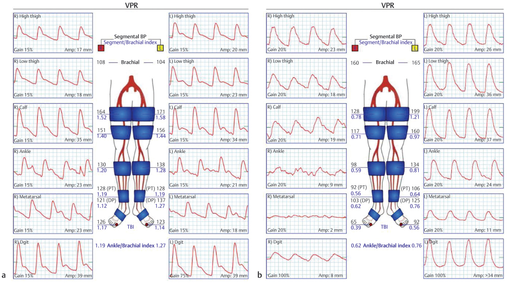
The ABI is a simple measurement that can be done with a sphygmomanometer and continuous wave Doppler probe. Brachial arterial systolic pressure serves as a reference for the determination of the ABI, giving an indication of the relative degree of ischemia. In addition, the absolute pressure measured at the ankle seems to correlate well with the prognosis of healing of skin wounds. Most surgeons would agree that absolute pressures less than 50 mm Hg signify rather severe PVD with a poor prognosis for healing.8,9 However, the ankle pressure measurement is not always accurate. Rigid arteries because of medial sclerosis, as most frequently seen in patients with diabetes, yield erroneously high-pressure measurements in the leg, giving falsely elevated ankle-brachial pressure indices. It is quite important, then, to obtain additional information with other noninvasive techniques, to assess the distal circulation more accurately. Patients with calcified arteries about the ankle should have measurements obtained from the great toe rather than the ankle vessels, as the toe digital arteries are less likely to have become calcified and can give a more reliable indication of pedal perfusion10 (▶ Fig. 3.1). Normal reference values for the ABI are shown in ▶ Table 3.2.
Pulse Volume Recordings
Pulse volume recordings provide a functional assessment of arterial perfusion of the lower extremity. PVRs use air plethysmography to determine changes in volume in the extremity during systole. Cuffs are placed at each segment of the extremity and inflated to a known pressure and volume. Pulse volume changes are measured as a change in pressure within the cuff and converted to an analog pressure pulse curve. Normal PVR waveforms have a systolic upstroke and a sharp systolic peak followed by a diastolic down stroke with a distinct dicrotic notch (▶ Fig. 3.1). Alterations in the pulse volume curve and amplitude suggest aberrant arterial flow with loss of the dicrotic notch and a widening of the complex, in mild to moderate disease, and eventual loss of amplitude in severe disease. PVRs are paired with segmental pressures and particularly helpful when extensive arterial calcinosis renders vessels noncompressible and gives falsely elevated ABIs. Waveforms are recorded from high in the thigh, the distal thigh, the calf, the ankle, and the great toe to detect and localize the region of the vascular disease.
Analog Doppler Waveforms
Though similar in many respects to a PVR, some patients, especially those with known PVD, benefit from the evaluation of Doppler velocity recordings. These are recorded from peripheral arteries and are important for the diagnosis and localization of occlusive PAD. The normal Doppler blood flow velocity waveform is triphasic, consisting of forward flow, reverse flow, and a second forward flow component (▶ Fig. 3.2a). Distal to an arterial stenosis or occlusion, the waveform is dampened, the amplitude of the velocity wave is decreased, the peak is delayed, and the reverse flow component is attenuated or absent (▶ Fig. 3.2b). In the usual vascular laboratory study, waveforms are recorded from the femoral, popliteal, dorsalis pedis, and posterior tibial arteries to detect and localize the region of the vascular disease. Although these studies provide only qualitative flow profiles, they assist both the vascular surgeon and plastic surgeon in planning their reconstructive approaches.
3.4.4 Imaging
Vascular imaging plays a crucial role in planning lower extremity reconstruction and provides important information about the patency of vessels, location of vascular lesions or obstructions, and the character of blood flow into the foot. Lower extremity angiography is the gold standard for diagnosing and characterizing the quality of arterial flow of the lower extremity. Angiography is an invasive procedure that must be done in a specialized room often with hybrid technology, making it expensive and cumbersome. Therefore, CT angiography (CTA), when available, has now supplanted conventional angiography in many cases. Atherosclerotic occlusive disease occurs and progresses in recognizable patterns. Lesions typically originate at the ostia of branch vessels or at main arterial bifurcations, and progress over time to affect multiple levels of the arterial tree. As a rule, younger patients will have isolated aortoiliac and focal superficial femoral arterial disease, whereas older patients may have occlusive disease that is more diffusely distributed. Patients with diabetes, however, are prone to isolated tibial and peroneal lesions rather than more proximal disease.11
Related posts:
 General Wound Preparation and Timing
 Supermicrosurgery Approach to the Lower Limb
 Modern Concepts of Prosthetic Rehabilitation in Amputation of the Lower Extremity
 Lower Limb Vascularized Composite Allotransplantation
 Using the Flap and Angiosome Concepts to Optimize Functional Lower Leg and Foot Amputations
 Procurement of Thin Flaps as Indicated in the Lower Extremity
Stay updated, free articles. Join our Telegram channel

Full access? Get Clinical Tree


