11 The Pertinence of the Reconstructive Ladder and the Reconstructive Elevator
Summary
If the goal in salvaging a lower limb were to just close the wound, then the stepwise approach beginning at the bottom rung of the reconstructive ladder that first chooses the simplest and least surgically morbid option would be considered, moving upward toward more complex alternatives only if unavoidable. Yet the reconstructive ladder is a flawed concept in that the goal initially should be instead the best long-term solution, and not just a solution. This can be best obtained by taking the reconstructive elevator to reach that floor where all the available choices can be scrutinized to allow an optimum selection process that will then obtain the desired durable, functional, as well as aesthetic outcome.
Keywords: reconstructive ladder, reconstructive elevator, long-term solution
11.1 Introduction
Descriptions and definitions of the reconstructive ladder permeate the literature and every discussion of reconstructive surgery. Medical students, surgical residents, and fellows are often still taught to approach wounds, traumatic injuries, and even congenital deformities within the framework of the reconstructive ladder (▶ Fig. 11.1). However, the reconstructive ladder is a flawed concept in that it implies that our reconstructive thought process should sequentially “go up” a ladder rather than directly to the best reconstructive option. The phrase reconstructive elevator was originally introduced to better describe the contemplative principles and practice of plastic surgery.1 There are few, if any, areas of plastic surgery where the power and implications of the reconstructive elevator are as pertinent as in lower extremity reconstruction.
The difference between a reconstructive ladder and a reconstructive elevator may seem to be a mere nuance of semantics. However, semantics are important. They shape our perceptions and frame our discussions. When used appropriately, they expand our horizons. When used inappropriately, they limit them. Remember that the best long-term solution, not just a solution, should then be that chosen initially. And that requires consideration of ALL options, any of which can be reached best by using the reconstructive elevator.
11.2 From Wound Closure to Reconstruction
To understand the flaws of the reconstructive ladder and the strengths of the reconstructive elevator, we must look at the origins of the terms. The reconstructive ladder is an improper extension of the well-known and appropriate concept of a wound-closure ladder. The wound-closure ladder signifies the stepwise algorithm used to decide on a wound-closure technique. It mandates starting with the simplest, least complex closure possibility, and then progress up the ladder only when required. For example, a clean, open wound that cannot be closed edge to edge would next be closed with a skin graft. If the wound bed would not support a skin graft, as if because of exposed bone without periosteum, then one would typically go up to the next rung of the wound-closure ladder and use a flap to close the wound.
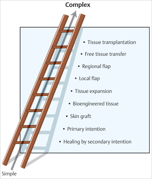
Fig. 11.1 The reconstructive ladder has a sequential approach to wound closure beginning with the simplest option adequate to close the wound starting at the bottom rung, then proceeding upward to more complex alternatives if indicated.
This concept of wound-closure ladder has its roots with the ancient Egyptians. The Smith Papyrus, written sometime between 2,600 and 200 BC, taught methods to treat different types of wounds. The papyrus advised that simple wounds should be “drawn together with ydr [suture].” Other wounds should be closed with “awy” strips with gum adhesive, while the infected wound cannot be closed immediately and should be treated with topical dressings including honey and sycamore leaves.2
The move from just wound closure to true reconstruction began in India around 600 BC when Sushruta described the reconstruction of noses with skin pedicled from the forehead—the first description of a technique that has evolved into the modern forehead flap. Their concern for preserving both form and function is linked to these principles of the ancient Indians, and marked the beginning of the modern era of plastic surgery. Where the concept of a wound-closure ladder is as accurate today as it was for the ancient Egyptians, the notion of a reconstructive ladder became obsolete with the ancient Indians and remains so.
The evolution of true lower extremity reconstruction then followed advances in the understanding of wound healing and the development of advanced surgical techniques. Due to its complex anatomy and wide spectrum of injuries, lower extremity reconstruction has lagged behind other areas of the body. Lower extremity amputation was the rule for most significant lower extremity wounds from the time of Hippocrates (460–370 BC) to that of Ambroise Pare (1509–1590). Ollier (1825–1900) developed the “occlusive technique” utilizing plaster of Paris for rest of the injured extremity. During the first World War, Winnett Orr combined the “closed plaster treatment” with incisional drainage of wounds. Joseph Trueta, in the Spanish Civil War (1935–1938), expanded on the closed treatment by performing surgical debridement of the wound prior to the placement of a plaster splint. Change was slow. Wounds were left to granulate and heal by secondary intention. Amputation rates may have decreased significantly, but postfracture osteomyelitis occurred in nearly 80% of patients. During World War II, the availability of antibiotics when added to Trueta technique decreased the rate of osteomyelitis to 25%.3
These historically poor results demonstrate the inadequacy of the reconstructive ladder in the lower extremity. Wounds of the lower extremity often involved and still do underlying fractures, or the exposure of vital structures in an area with a paucity of redundant vascular soft tissue. While the wounds may have been “closed” with the Trueta approach, the final result was often less than satisfactory. Early methods to compensate for inadequate soft tissues included the cross-leg flap described by Hamilton and tubed pedicle flaps by Gilles. Yet these techniques were time-consuming, required multiple operations, and often resulted in joint contractures and inhibited ambulatory function that as an option may have been no better.
11.3 The Modern Era of Lower Extremity Reconstruction
It was not until the discovery of regional lower extremity flaps in the 1960s that true reconstruction of the lower extremity became practical. The development of microsurgery in the 1970s, reintroduction of lower extremity fasciocutaneous flaps in the 1980s,4 and expansion of the angiosome concept5,6 that led to perforator free flaps and local free-style perforator flaps7,8,9,10,11 increased the options for flaps while decreasing donor site morbidity. Suprafascial thin adipocutaneous flaps and perforator-to-perforator free tissue transfer utilizing supermicrosurgical techniques are the ultimate extension of this experience.12,13 In many cases, these now established techniques allow for the closure of lower extremity wounds that may otherwise have required amputation.
With the advent of local perforator flaps, and perforator-to-perforator free flaps, one could say that more rungs have been added to the reconstructive ladder. However, this advancement is more appropriately viewed as an adoption of parallel thinking required within the reconstructive elevator (▶ Fig. 11.2). When the goal is not to just close a wound but also to maximize form and function and minimize donor-site morbidity, pick the best floor possible by taking the reconstructive elevator to choose which of the similar yet different options would be the more suitable.
11.4 The Reconstructive Elevator and the Lower Extremity
To reiterate in this era when form and function must be the goal, the concept of the reconstructive elevator implies that simplest is not necessarily the best. To think sequentially about what is the next better reconstruction option no longer suffices. Modern technology and advanced techniques have made such thinking obsolete. Reconstructive surgery as we now know it requires parallel, creative thought processes rather than simple sequential thinking. The reconstructive surgeon must be capable of skipping a rung of the ladder and take the elevator up to the next floor or two when necessary.
When approaching a challenging lower extremity defect using the concept of the reconstructive elevator, the most important step for the surgeon is to consider the outcome possible due to surgical intervention for that specific patient. Is the goal of the surgery to get the wound closed with minimal additional morbidity or anesthetic time for the patient? Is the goal of the surgery to close the wound maximizing the aesthetic and functional result for the patient? Is the goal of the surgery to close the wound and reconstruct other vital structures such as tendon, bone, or nerve? The answers to these questions often depend not only on the characteristics of the wound, but also on factors specific to the patient such as their anesthetic risk, potential functional status, and need for future procedures or postoperative adjunctive therapy such as radiation.
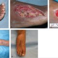
Sometimes clinical examples will better clarify these points. Consider the case of a patient with a soft-tissue defect after tumor extirpation (▶ Fig. 11.3). If the only goal were to close the wound, the Trueta technique of letting it heal by secondary intention may be adequate. Alternatively, a skin graft could be used to shorten the healing time with less risk of scar contracture. However, if the goal is to not only close the wound but also to provide long-term stable coverage that best tolerates the shear forces that constantly impinge on the lower limb, restore an optimal soft-tissue contour, and minimize any potential of joint contracture, then it would be appropriate to skip the lower rungs of the ladder and jump on the elevator to proceed with the best flap coverage.
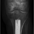
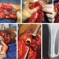
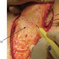
Consider also a patient who had an injury resulting in both a segmental loss of the distal Achilles tendon and the overlying soft tissue. A sequential plan using the reconstructive ladder might indicate a reverse sural flap to close the wound, but without tendon reconstruction, to be the most appropriate next step. This, however, could result in life-long use of an ankle foot orthotic or multiple secondary operations to reconstruct the Achilles tendon. Alternatively, one could jump on the elevator and both close the wound and reconstruct the Achilles tendon in a single stage using a conjoined anterolateral thigh free flap along with vascularized fascia lata (▶ Fig. 11.4, ▶ Fig. 11.5). In these examples, prioritizing both form and function necessitated choosing the better option, even if a simpler yet less ideal option was feasible.

Related posts:
 General Wound Preparation and Timing
 Supermicrosurgery Approach to the Lower Limb
 Modern Concepts of Prosthetic Rehabilitation in Amputation of the Lower Extremity
 Lower Limb Vascularized Composite Allotransplantation
 Using the Flap and Angiosome Concepts to Optimize Functional Lower Leg and Foot Amputations
 Procurement of Thin Flaps as Indicated in the Lower Extremity
Stay updated, free articles. Join our Telegram channel

Full access? Get Clinical Tree


